

Cytokine profile during latent and slowly progressive primary tuberculosis: a possible role for interleukin-15 in mediating clinical disease
- PMID: 16367949
- PMCID: PMC1809553
- DOI: 10.1111/j.1365-2249.2005.02976.x
Cytokine profile during latent and slowly progressive primary tuberculosis: a possible role for interleukin-15 in mediating clinical disease
Abstract
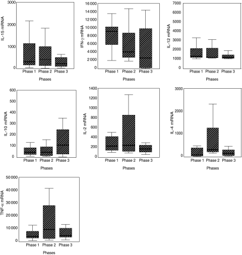
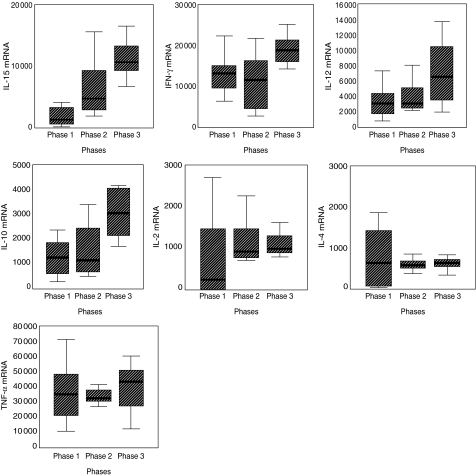
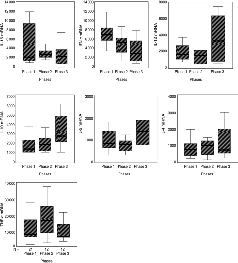
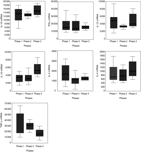
Recently, mouse models for latent (LTB) and slowly progressive primary tuberculosis (SPTB) have been established. However, cytokine profiles during the two models are not well established. Using quantitative reverse transcription-polymerase chain reaction (qRT-PCR) we studied the expression levels of interleukin (IL)-2, IL-4, IL-10, IL-12, IL-15, interferon (IFN)-gamma and tumour necrosis factor (TNF)-alpha during the course of LTB and SPTB in the lungs and spleens of B6D2F1Bom mice infected with the H37Rv strain of Mycobacterium tuberculosis (Mtb). The results show that, except for IL-4, cytokine expression levels were significantly higher during SPTB than LTB in both the lungs and spleens. During LTB, all the cytokines (except IL-2 in the lungs) had higher expression levels during the initial period of infection both in the lungs and spleens. During SPTB, the expression levels of IL-15 increased significantly from phases 1 to 3 in the lungs. The expression levels of IL-10, IL-12 and IFN-gamma increased significantly from 2 to 3 in the lungs. IL-10 and IL-15 increased significantly from phases 2 to 3, whereas that of TNF-alpha decreased significantly and progressively from phases 1 to 3 in the spleens. Over-expression of proinflammatory cytokines during active disease has been well documented, but factor(s) underlying such over-expression is not known. In the present study, there was a progressive and significant increase in the expression levels of IL-15, together with Th1 cytokines (IL-12 and IFN-gamma) during SPTB but a significant decrease during LTB. IL-15 is known to up-regulate the production of proinflammatory cytokines, IL-1beta, IL-8, IL-12, IL-17, IFN-gamma and TNF-alpha and has an inhibitory effect on activation-induced cell death. IL-15 is known to be involved in many proinflammatory disease states such as rheumatoid arthritis, sarcoidosis, inflammatory bowel diseases, autoimmune diabetes, etc. Our results, together with the above observations, suggest that IL-15 may play an important role in mediating active disease during Mtb infection.
Figures
Similar articles
-
Differential regulation of Th1-type and Th2-type cytokine profiles in pancreatic islets of C57BL/6 and BALB/c mice by multiple low doses of streptozotocin.Immunobiology. 2002 Mar;205(1):35-50. doi: 10.1078/0171-2985-00109. Immunobiology. 2002. PMID: 11999343
-
Sex-determined susceptibility and differential IFN-gamma and TNF-alpha mRNA expression in DBA/2 mice infected with Leishmania mexicana.Immunology. 1995 Jan;84(1):1-4. Immunology. 1995. PMID: 7890293 Free PMC article.
-
Intragraft cytokine mRNA expression in rejecting and non-rejecting vascularized xenografts.Xenotransplantation. 2003 Jul;10(4):311-24. doi: 10.1034/j.1399-3089.2003.02032.x. Xenotransplantation. 2003. PMID: 12795680
-
The role of cytokine mRNA stability in the pathogenesis of autoimmune disease.Autoimmun Rev. 2006 May;5(5):299-305. doi: 10.1016/j.autrev.2005.10.013. Epub 2005 Dec 21. Autoimmun Rev. 2006. PMID: 16782553 Review.
-
Immunostimulatory cytokines in somatic cells and gene therapy of cancer.Cytokine Growth Factor Rev. 1997 Jun;8(2):119-28. doi: 10.1016/s1359-6101(96)00052-4. Cytokine Growth Factor Rev. 1997. PMID: 9244407 Review.
Cited by
-
Mouse lung and spleen natural killer cells have phenotypic and functional differences, in part influenced by macrophages.PLoS One. 2012;7(12):e51230. doi: 10.1371/journal.pone.0051230. Epub 2012 Dec 5. PLoS One. 2012. PMID: 23227255 Free PMC article.
-
Review: Impact of Helminth Infection on Antimycobacterial Immunity-A Focus on the Macrophage.Front Immunol. 2017 Dec 22;8:1864. doi: 10.3389/fimmu.2017.01864. eCollection 2017. Front Immunol. 2017. PMID: 29312343 Free PMC article. Review.
-
Association of ESAT-6/CFP-10-induced IFN-γ, TNF-α and IL-10 with clinical tuberculosis: evidence from cohorts of pulmonary tuberculosis patients, household contacts and community controls in an endemic setting.Clin Exp Immunol. 2017 Aug;189(2):241-249. doi: 10.1111/cei.12972. Epub 2017 May 2. Clin Exp Immunol. 2017. PMID: 28374535 Free PMC article.
-
The emergence of Beijing family genotypes of Mycobacterium tuberculosis and low-level protection by bacille Calmette-Guérin (BCG) vaccines: is there a link?Clin Exp Immunol. 2006 Sep;145(3):389-97. doi: 10.1111/j.1365-2249.2006.03162.x. Clin Exp Immunol. 2006. PMID: 16907905 Free PMC article. Review.
-
Characterization of peripheral cytokine-secreting cells responses in HIV/TB co-infection.Front Cell Infect Microbiol. 2023 Jul 6;13:1162420. doi: 10.3389/fcimb.2023.1162420. eCollection 2023. Front Cell Infect Microbiol. 2023. PMID: 37483385 Free PMC article.
References
-
- World Health Organization (WHO). The world health report, global tuberculosis control. Geneva: WHO; 2001. Report no. 287.
-
- Taha RA, Kotsimbos TC, Song YL, Menzies D, Hamid Q. IFN-gamma and IL-12 are increased in active compared with inactive tuberculosis. Am J Respir Crit Care Med. 1997;155:1135–9. - PubMed
-
- Grabstein KH, Eiseman J, Shanebeck K, et al. Cloning of a T cell growth factor that interacts with the beta chain of interleukin-2 receptor. Science. 1994;264:965–8. - PubMed
MeSH terms
Substances
LinkOut - more resources
Full Text Sources
Medical
