

Packaging of actin into Ebola virus VLPs
- PMID: 16367999
- PMCID: PMC1334228
- DOI: 10.1186/1743-422X-2-92
Packaging of actin into Ebola virus VLPs
Abstract
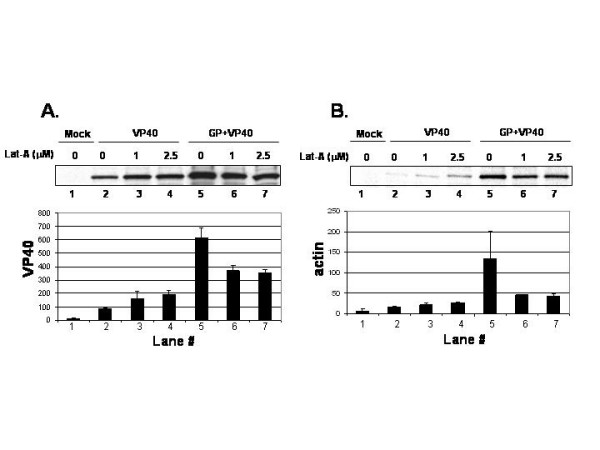
The actin cytoskeleton has been implicated in playing an important role assembly and budding of several RNA virus families including retroviruses and paramyxoviruses. In this report, we sought to determine whether actin is incorporated into Ebola VLPs, and thus may play a role in assembly and/or budding of Ebola virus. Our results indicated that actin and Ebola virus VP40 strongly co-localized in transfected cells as determined by confocal microscopy. In addition, actin was packaged into budding VP40 VLPs as determined by a functional budding assay and protease protection assay. Co-expression of a membrane-anchored form of Ebola virus GP enhanced the release of both VP40 and actin in VLPs. Lastly, disruption of the actin cytoskeleton with latrunculin-A suggests that actin may play a functional role in budding of VP40/GP VLPs. These data suggest that VP40 may interact with cellular actin, and that actin may play a role in assembly and/or budding of Ebola VLPs.
Figures

Similar articles
-
Effect of Ebola virus proteins GP, NP and VP35 on VP40 VLP morphology.Virol J. 2006 May 23;3:31. doi: 10.1186/1743-422X-3-31. Virol J. 2006. PMID: 16719918 Free PMC article.
-
Ebola virus VP35-VP40 interaction is sufficient for packaging 3E-5E minigenome RNA into virus-like particles.J Virol. 2006 Jun;80(11):5135-44. doi: 10.1128/JVI.01857-05. J Virol. 2006. PMID: 16698994 Free PMC article.
-
Overlapping motifs (PTAP and PPEY) within the Ebola virus VP40 protein function independently as late budding domains: involvement of host proteins TSG101 and VPS-4.J Virol. 2003 Feb;77(3):1812-9. doi: 10.1128/jvi.77.3.1812-1819.2003. J Virol. 2003. PMID: 12525615 Free PMC article.
-
Conformational plasticity of the Ebola virus matrix protein.Protein Sci. 2014 Nov;23(11):1519-27. doi: 10.1002/pro.2541. Epub 2014 Sep 4. Protein Sci. 2014. PMID: 25159197 Free PMC article. Review.
-
Structural studies on the Ebola virus matrix protein VP40 indicate that matrix proteins of enveloped RNA viruses are analogues but not homologues.FEMS Microbiol Lett. 2004 Apr 15;233(2):179-86. doi: 10.1016/j.femsle.2004.03.002. FEMS Microbiol Lett. 2004. PMID: 15108720 Free PMC article. Review.
Cited by
-
An Ebola Virus-Like Particle-Based Reporter System Enables Evaluation of Antiviral Drugs In Vivo under Non-Biosafety Level 4 Conditions.J Virol. 2016 Sep 12;90(19):8720-8. doi: 10.1128/JVI.01239-16. Print 2016 Oct 1. J Virol. 2016. PMID: 27440895 Free PMC article.
-
A cationic, C-terminal patch and structural rearrangements in Ebola virus matrix VP40 protein control its interactions with phosphatidylserine.J Biol Chem. 2018 Mar 2;293(9):3335-3349. doi: 10.1074/jbc.M117.816280. Epub 2018 Jan 18. J Biol Chem. 2018. PMID: 29348171 Free PMC article.
-
Bimolecular Complementation to Visualize Filovirus VP40-Host Complexes in Live Mammalian Cells: Toward the Identification of Budding Inhibitors.Adv Virol. 2011 Jan 1;2011:341816. doi: 10.1155/2011/341816. Adv Virol. 2011. PMID: 22102845 Free PMC article.
-
Host Protein BAG3 is a Negative Regulator of Lassa VLP Egress.Diseases. 2018 Jul 13;6(3):64. doi: 10.3390/diseases6030064. Diseases. 2018. PMID: 30011814 Free PMC article.
-
Predictive and comparative analysis of Ebolavirus proteins.Cell Cycle. 2015;14(17):2785-97. doi: 10.1080/15384101.2015.1068472. Epub 2015 Jul 9. Cell Cycle. 2015. PMID: 26158395 Free PMC article.
References
-
- Timmins J, Scianimanico S, Schoehn G, Weissenhorn W. Vesicular release of ebola virus matrix protein VP40. Virology. 2001;283:1–6. - PubMed
Publication types
MeSH terms
Substances
Grants and funding
LinkOut - more resources
Full Text Sources
