. 2005 Dec 20:6:30.
doi: 10.1186/1471-2091-6-30.

Computer simulation of partitioning of ten pentapeptides Ace-WLXLL at the cyclohexane/water and phospholipid/water interfaces

Affiliations

Computer simulation of partitioning of ten pentapeptides Ace-WLXLL at the cyclohexane/water and phospholipid/water interfaces

Marcela P Aliste et al. BMC Biochem. .

Abstract

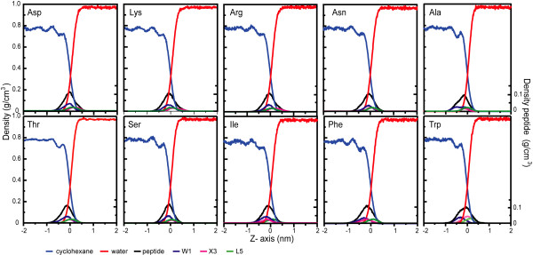
Background: Peptide-membrane interactions play a key role in the binding, partitioning and folding of membrane proteins, the activity of antimicrobial and fusion peptides, and a number of other processes. To gain a better understanding of the thermodynamics of such interactions, White and Wimley created an interfacial hydrophobicity scale based of the transfer free energy from water to octanol or lipid bilayers of a series of synthetic peptapeptides (Ace-WLXLL, with X being any of the twenty natural amino acids) (White and Wimley (1996) Nat. Struct. Biol. 3, 842-848). In this study, we performed molecular dynamics simulations of a representative set of ten of these peptides (X = D, K, R, N, A, T, S, I, F and W) in two membrane mimetic interfaces: water-cyclohexane (10 ns) and a fully solvated dioleoylphosphatidylcholine (DOPC) bilayer (50 ns) using both constant pressure and constant area ensembles. We focus on partitioning of the ten peptides at the cyclohexane/water and lipid/water interfaces.

Results: The peptides rapidly equilibrate (< 2 ns) and partition at the cyclohexane/water interface. The X3 guest residue assumes average orientations that depend on the nature of the side chain. At the DOPC/water interface, dynamics is much slower and convergence is difficult to achieve on a 50 ns timescale. Nonetheless, all peptides partition to the lipid/water interface with distributions with widths of 1-2 nm. The peptides assume a broad range of side chain and backbone orientations and have only a small effect on the area of the unit cell. On average, hydrophobic guest residues partition deeper into the hydrophobic core than hydrophilic residues. In some cases the peptides penetrate sufficiently deep to somewhat affect the distribution of the C=C double bond in DOPC. The relative distribution of the X3 guest residue compared to W1 and L5 is similar in the water/cyclohexane and water/lipid simulations. Snapshots show mostly extended backbone conformations in both environments. There is little difference between simulations at a constant area of 0.66 nm2 and simulations at constant pressure that approximately yield the same average area of 0.66 nm2.

Conclusion: These peptides were designed to assume extended conformations, which is confirmed by the simulations. The distribution of the X3 side chain depends on its nature, and can be determined from molecular dynamics simulations. The time scale of peptide motion at a phospholipids-water interface is too long to directly calculate the experimentally measured hydrophobicity scale to test and improve the simulation parameters. This should be possible at the water/cyclohexane interface and likely will become feasible in the future for the phospholipids/water case.


Figures

Figure 1
Figure 1
Overview of the simulation systems. Starting configurations: (a) the water/cyclohexane system and (b) the DOPC bilayer. Phosphorous atoms are shown in orange, nitrogen atoms in blue. The peptides are shown in a space-filling representation.
Figure 2
Figure 2
Snapshots of the ten peptides in the water/cyclohexane system after 10 ns. The peptide backbone is violet, the Ace and C-terminus are colored by atom type, Trp is green, Leu cyan, and the changing residue X3 is orange, while the water is shown as small white spheres and the cyclohexane is shown in light grey.
Figure 3
Figure 3
The z-coordinates of the center of mass of the entire peptide, three side chains, water and cyclohexane as function of time for all ten cyclohexane/water systems. Trajectory profiles along the z-coordinates for the peptides in the cyclohexane/water systems. The zeropoint is the point where the water and cyclohexane densities are equal (see figure 4).
Figure 4
Figure 4
Density profiles along the z-axis. The density profiles of the peptides have been averaged over the last 2 ns of simulation. The scales on the left and right axes correspond to the solvent components (water and cyclohexane) and peptides and/or side chains, respectively. The middle of the interface (0 nm) is defined as the point where the partial density of the water equals the partial density of the cyclohexane.
Figure 5
Figure 5
A. Snapshots of all peptides except the Trp peptide in the DOPC lipid bilayer system at 50 ns (NpT simulations). The first row and the third represent the peptide in the upper leaflet; the second and the last row the peptide in the lower leaflet. The peptide backbone is violet, the Ace and C-terminus are colored by atom type, Trp is green, Leu cyan, and the changing residue X3 is orange, while the water is shown as small white spheres and the cyclohexane is shown in light grey. The water molecules are shown as white spheres. The DOPC lipid bilayer is shown as two separated groups: the head group (choline and phosphate) is dark grey while the tails are lighter gray.
Figure 6
Figure 6
A close-up of the Trp peptide, in the same representation as the other 9 peptides in Figure 5.
Figure 7
Figure 7
Trajectory profiles along the z-coordinates for the peptides in the lipid bilayer system (NpAT simulations). The z-coordinates for the center of mass of the different groups are plotted averaged over 10 ps. The groups are the two peptides, the three side chains in positions Trp1, X3 and Leu5 with respect to the average center of mass (0 nm) of the DOPC group. The lipid bilayer interface is defined with the coordinates: the choline group (N) and the first CH2 group of the lipid chain (the second carbon counting from the ester link, C2).
Figure 8
Figure 8
Trajectory profiles along the z-coordinates for the peptides in the lipid bilayer system (NpT simulations). The z coordinates for the center of mass of the different groups: peptide, three side chains in positions Trp1, X3 and Leu5 with respect to the average center of mass (0 nm) of the DOPC group. The lipid bilayer interface is defined with the coordinates of the choline group (N) and the first CH2 group of the lipid chain (the second carbon counting from the ester link, C2).
Figure 9
Figure 9
Density profiles along the bilayer normal (NpAT simulations). The profiles were obtained over the last 10 ns of the simulations; the left and right scales of the graph correspond to the lipid components and the peptides and/or side chain, respectively. Partial density for some of the interface components of the lipid bilayers (as choline, phosphate and carbonyl) and one component of the hydrocarbon core of the bilayers, the double bond distribution. Partial densities of the peptides are show as solid representation and the three side chains are shown Trp1, X3 and Leu5.
Figure 10
Figure 10
Density profiles along the bilayer normal (NpT simulations). The profiles were obtained over the last 10 ns of the simulations; the left and right scales of the graph correspond to the lipid components and the peptides and/or side chain, respectively. Partial density for some of the interface components of the lipid bilayers (as choline, phosphate and carbonyl) and one component of the hydrocarbon core of the bilayers, the double bond distribution. Partial densities of the peptides are show as solid representation and the three side chains are shown Trp1, X3 and Leu5.
Figure 11
Figure 11
Orientations of the Trp1 ring (NpT simulations) as a function of time, defined by SL and SN.

References

    1. Kessel A, Ben-Tal N. Free energy determinants of peptide association with lipids bilayers. Current Topics in Membranes. 2002;52:205–253.
    1. Epand RM. Fusion peptides and the mechanism of viral fusion. Biochim Biophys Acta. 2003;1614:116–121. - PubMed
    1. Epand RM, Vogel HJ. Diversity of antimicrobial peptides and their mechanisms of action. Biochim Biophys Acta. 1999;1462:11–28. - PubMed
    1. Murray D, Arbuzova A, Honig B, MacLaughlin S. The role of electrostatic and nonpolar interactions in the association of peripheral protiens with membranes. Peptide-lipid interactions. 2002;52:277–307.
    1. Wimley WC, White SH. Experimentally determined hydrophobicity scale for proteins at membrane interfaces. Nat Struct Biol. 1996;3:842–848. - PubMed

Publication types