Review
. 2006 Jan;133(2):183-93.
doi: 10.1242/dev.02214.

Shaping BMP morphogen gradients in the Drosophila embryo and pupal wing

Affiliations
Review

Shaping BMP morphogen gradients in the Drosophila embryo and pupal wing

Michael B O'Connor et al. Development. 2006 Jan.

Abstract

In the early Drosophila embryo, BMP-type ligands act as morphogens to suppress neural induction and to specify the formation of dorsal ectoderm and amnioserosa. Likewise, during pupal wing development, BMPs help to specify vein versus intervein cell fate. Here, we review recent data suggesting that these two processes use a related set of extracellular factors, positive feedback, and BMP heterodimer formation to achieve peak levels of signaling in spatially restricted patterns. Because these signaling pathway components are all conserved, these observations should shed light on how BMP signaling is modulated in vertebrate development.


Figures

Fig. 1.
Fig. 1.. BMP signaling in Drosophila.
Three different BMP ligand species exist in the early embryo. (A) Dpp homodimers, (B) Dpp/Scw heterodimers, and (C) Scw homodimers. (B) Dpp/Scw heterodimers are preferentially transported to the midline through the action of Sog/Tsg and Tld. At the midline, the heterodimer accumulates and is free for signaling as Tld processes Sog. This heterodimer binds to a heteromeric receptor complex, probably a tetramer located in the plasma membrane (PM), composed of two type II receptors (Punt), and one subunit each of the type I receptors Tkv and Sax. Punt activates Tkv and Sax by phosphorylating residues within their GS boxes (a glycine-serine rich segment near the membrane). Once activated, the type I receptors phosphorylate Mad. Mad then associates with the co-Smad Medea (probably in a trimeric complex of uncharacterized subunit composition), and the complex translocates to the nucleus where it binds to and activates or represses target genes in conjunction with other transcription factors (TFs). At the midline, the Sax and Tkv receptors produce a synergistic signal that results in the activation of high-threshold target genes, such as race. In the lateral regions, homodimers of Scw and Dpp produce moderate and low levels signals, respectively, that can activate low-threshold response genes, such as pannier (pnr) (for details, see Shimmi et al., 2005b).
Fig. 2.
Fig. 2.. Patterning the dorsal side of the Drosophila embryo by BMP transport.
(A) Dpp is transcribed uniformly within the dorsal half of the embryo in the early blastoderm. (B) Initially, Mad phosphorylation is wide and of low intensity at the mid-cellular stage, but then refines during late blastoderm into a sharper and more intense stripe. The refinement requires an additional unknown factor that is induced by the early low-level Dpp signal (see Wang and Ferguson, 2005). (C) A schematic cross-sectional representation of an embryo showing the expression domains of the various extracellular components (red, sog; blue, tsg and tld; green, area of overlap in dpp and scw expression). Sog diffuses into the dorsal domain from its ventrolateral site of synthesis, and preferentially complexes with Dpp/Scw heterodimers and Tsg. (D) Net diffusion of this complex, driven in part by Tld processing of Sog, promotes accumulation of the Dpp/Scw heterodimer near the midline from mid- to late-cellular blastoderm stage. Homodimers of Dpp and Scw are not transported efficiently as they have a lower affinity for the Sog/Tsg complex (see Shimmi et al., 2005b). (E) The spatial distribution of BMP-bound receptor at various times obtained using modeling methods similar to those described by Mizutani et al. (Mizutani et al., 2005) in which there is a constant BMP production/degradation. Note that the model predicts that, at a given threshold, the intensity of the pMad stripe should both increase in time and widen. NE, neuroectoderm; DM, dorsal midline.
Fig. 3.
Fig. 3.. Posterior crossvein formation requires BMPs and BMP transport components.
(A,B) pMad accumulation in the longitudinal veins (LVs) and posterior crossvein (PCV) at 26 (A) and 36 (B) hours post-puparium formation (ppf). Note that at 26 hours ppf pMad accumulates at the PCV in a wide domain that then refines considerably by 36 hours ppf. (C) dpp mRNA expression in the LVs, but not in the PCV, at 24 hours ppf. (D) cv-2 mRNA expression at 24 and 29 hours ppf. Note the sharpening in the cv-2 mRNA profile as time progresses. (E) A schematic representation of one possible patterning mechanism. Dpp is only produced in the LVs, whereas Gbb is uniformly expressed. The Dpp/Gbb heterodimers formed in the LVs preferentially bind to a complex of Sog and Cv (also known as Tsg2). Tlr cleaves Sog to release the heterodimer for signaling. Initial low signal levels, together with other unknown positional cues, induce cv-2 transcription (yellow) in a zone that will form the PCV. Cv-2 protein accumulates on the cell surface and creates a positive-feedback loop that presents BMP ligand to the signaling receptors. (F-I) Expression patterns of sog (F), cv (G) and tlr (H) mRNA, and Tkv protein (I) in 19–24 hour ppf wings. (J) Uniform overexpression of sog (UAS-sog), cv (EP(X)1349) and cv-2 (EP(2)1103) in the posterior of the wing with en-gal4 does not disrupt PCV formation. (C,F,I) Reproduced, with permission, from Ralston and Blair (Ralston and Blair, 2005); (D,H,J) reproduced, with permission, from Ralston (A. Ralston, PhD thesis, University of Wisconsin, 2004); (G) reproduced, with permission, from Shimmi et al. (Shimmi et al., 2005a).
Fig. 4.
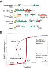
Fig. 4.. CR-containing proteins and their possible roles in positive feedback and spatial bi-stability.
(A) The domain structures of several BMP-binding proteins. Sog is a secreted (signal peptide, SP, yellow) protein that contains BMP-binding modules [cysteine-rich motifs (CRs), green], whereas Cv-2, and its vertebrate homologs (not shown), and other related vertebrate proteins, such as Kielin or KCP, contain both CR domains and a von Willebrand Factor D (VWFD) motif (red) that may promote cell surface localization. The CRIM1 protein also contains CR domains but instead of a VWFD, it contains an insulin-like growth factor binding protein domain (IGFBP), a transmembrane domain (TM) that is likely to anchor it to the cell surface, and a small cytoplasmic tail (Cyto). (B) Position (or BMP) versus BMP-bound receptor for on/off equilibrium (dotted line) and positive feedback induced bi-stability (solid line). In general the distribution of morphogen is non-linear in x [BMP=f(x)]; however, in this case, the extracellular gradient of BMP is linear in position (i.e. BMP~α*x). As the level of BMP increases, the level of BMP-bound receptor follows the on/off equilibrium solution until it reaches a limit point (LP) where the lower equilibrium solution ceases to exist. For levels of BMP above this point, the level of BMP-bound receptor approaches the upper stable branch.
Fig. 5.
Fig. 5.. A model for positive feedback.
Initially BMP ligands, such as Dpp and Scw, bind to the type I and type II signaling receptors. This signal activates transcription of a cell surface BMP-binding protein (sbp), such as Cv-2, that helps to present ligand to the signaling receptors. This may account for the production of spatial bi-stability, as proposed by Ferguson and Wang (Ferguson and Wang, 2005). P, phosphorylation. The question mark indicates that the identity of this component is not yet established in the embryo.

References

    1. Aono A, Hazama M, Notoya K, Taketomi S, Yamasaki H, Tsukuda R, Sasaki S and Fujisawa Y (1995). Potent ectopic bone-inducing activity of bone morphogenetic protein-4/7 heterodimer. Biochem. Biophys. Res. Commun 210, 670–677. - PubMed
    1. Araujo H, Negreiros E and Bier E (2003). Integrins modulate Sog activity in the Drosophila wing. Development 130, 3851–3864. - PubMed
    1. Arora K and Nusslein-Volhard C (1992). Altered mitotic domains reveal fate map changes in Drosophila embryos mutant for zygotic dorsoventral patterning genes. Development 114, 1003–1024. - PubMed
    1. Arora K, Levine MS and O’Connor MB (1994). The screw gene encodes a ubiquitously expressed member of the TGF-beta family required for specification of dorsal cell fates in the Drosophila embryo. Genes Dev. 8, 2588–2601. - PubMed
    1. Ashe HL and Levine M (1999). Local inhibition and long-range enhancement of Dpp signal transduction by Sog. Nature 398, 427–431. - PubMed

Publication types

MeSH terms

LinkOut - more resources