. 2006 Jan;74(1):88-98.
doi: 10.1128/IAI.74.1.88-98.2006.

Dissection of ESAT-6 system 1 of Mycobacterium tuberculosis and impact on immunogenicity and virulence

Affiliations
Free PMC article

Dissection of ESAT-6 system 1 of Mycobacterium tuberculosis and impact on immunogenicity and virulence

Priscille Brodin et al. Infect Immun. 2006 Jan.
Free PMC article

Abstract

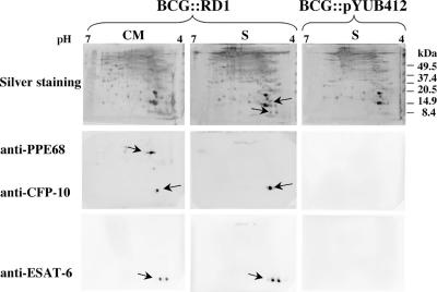
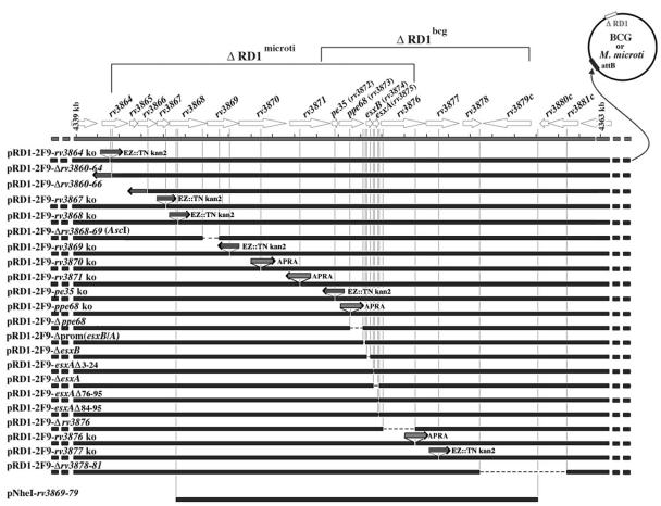
The dedicated secretion system ESX-1 of Mycobacterium tuberculosis encoded by the extended RD1 region (extRD1) assures export of the ESAT-6 protein and its partner, the 10-kDa culture filtrate protein CFP-10, and is missing from the vaccine strains M. bovis BCG and M. microti. Here, we systematically investigated the involvement of each individual ESX-1 gene in the secretion of both antigens, specific immunogenicity, and virulence. ESX-1-complemented BCG and M. microti strains were more efficiently engulfed by bone-marrow-derived macrophages than controls, and this may account for the enhanced in vivo growth of ESX-1-carrying strains. Inactivation of gene pe35 (Rv3872) impaired expression of CFP-10 and ESAT-6, suggesting a role in regulation. Genes Rv3868, Rv3869, Rv3870, Rv3871, and Rv3877 encoding an ATP-dependent chaperone and translocon were essential for secretion of ESAT-6 and CFP-10 in contrast to ppe68 Rv3873 and Rv3876, whose inactivation did not impair secretion of ESAT-6. A strict correlation was found between ESAT-6 export and the generation of ESAT-6 specific T-cell responses in mice. Furthermore, ESAT-6 secretion and specific immunogenicity were almost always correlated with enhanced virulence in the SCID mouse model. Only loss of Rv3865 and part of Rv3866 did not affect ESAT-6 secretion or immunogenicity but led to attenuation. This suggests that Rv3865/66 represent a new virulence factor that is independent from ESAT-6 secretion. The present study has allowed us to identify new aspects of the extRD1 region of M. tuberculosis and to explore its role in the pathogenesis of tuberculosis.


Figures

FIG. 1.
FIG. 1.
Two-dimensional analysis of whole-cell extracts (CM) and supernatants (S) of BCG::RD1 and control BCG::pYUB412. Detection was carried out by silver staining (top) and with monoclonal anti-ESAT-6 and polyclonal anti-CFP-10 and anti-PPE68 antibodies for Western blot analysis.
FIG. 2.
FIG. 2.
(A) Quantification of bacterial uptake (%) at 16 h postinfection of murine bone marrow macrophages (BMMΦ) with BCG and M. microti expressing the whole RD1 region (+RD1) or not (−) in the presence or absence of Tween 80. CFU in BMMΦ (B) or after activation with LPS and IFN-γ (C) at 4 h (□) and days 3 (░⃞) and 6 (▪) after infection with BCG::pYUB412, BCG::RD1, and M. tuberculosis H37Rv. Means and standard deviations are from infections done in quadruplicate wells and are representative of four experiments.
FIG. 3.
FIG. 3.
In vivo bacterial replication of BCG::RD1 in various mouse models: C57BL/6 wild-type mice (open bars), B6;129-LT-α/TNF-α ko mice (light gray bars), inbred 129/Sv IFN-γ receptor ko mice (dark gray bars), and SCID mice (black bars). The ratios of CFU counts at day 28 in spleen (A) and lungs (B) relative to initial dose after intravenous infection with 106 CFU are indicated. Each value is the mean of three or four mice, and the error bars represent standard deviations. (C) Histological samples of lungs from C57BL/6 wild type (WT), TNF-α ko, and IFN-γ receptor ko mice, showing compact discrete lesions in lungs of C57BL/6 mice and large diffuse lesions for TNF-α ko mice infected with BCG::RD1 compared to the absence of lesions in wild-type and TNF-α ko mice infected with the BCG vector control. Lungs from IFN-γ receptor ko mice showed lesions with both strains, although bacterial counts were lower for the vector control. Sections were stained with hematoxylin and eosin. Magnification, ×100.
FIG. 4.
FIG. 4.
Scheme of the various pRD1-2F9 and pNheI constructs that were integrated into BCG and M. microti. Within the genome shown as a circle, ΔRD1 refers to the naturally occurring RD1 deletions in M. microti and BCG. For each construct (the name is given above on the left), the site of the transposon insertion (EZ::TN kan2), apramycin cassette (APRA), or deletion (—) in the corresponding open reading frames relative to its position in RD1 region is shown. The orientation of the transposon/cassette is shown by the direction of the arrow. pe35, ppe68, esxB, and esxA correspond to Rv3872, Rv3873, Rv3874, and Rv3875.
FIG. 5.
FIG. 5.
Secretion analysis. In vitro expression and secretion of ESX-1 antigens from recombinant M. microti, BCG, and M. tuberculosis H37RvΔRD1 complemented with integrating cosmids that were mutated in selected RD1 genes was examined. Total protein concentrations were determined by using Bio-Rad protein assay, and 20-μg samples were subjected to SDS-PAGE. PPE68, CFP-10, and ESAT-6 correspond to products encoded by ppe68 (Rv3873), esxB (Rv3874), and esxA (Rv3875) genes, respectively. (A) CM, whole-cell extract containing cytosolic and membrane fractions; S, supernatant. Detection was carried out by using monoclonal anti-ESAT-6, polyclonal anti-CFP-10, and polyclonal anti-PPE68 antibodies. (B) Analysis of recombinant M. tuberculosis pe35 ko and control strains: (a) secretion analysis with same antibodies as described for panel A; (b) PCR control amplification obtained with primers for genes pe35 to Rv3877 for pe35 ko and RD1-2F9 M. tuberculosis H37RvΔRD1 strains; (c) hybridization signals obtained with radiolabeled cDNA from M. tuberculosis H37RvΔRD1::RD1-2F9 and pe35 ko mutant using a focused mini-array containing PCR products from selected genes of the RD1 region spotted on nylon membranes in duplicates. (C) Secretion analysis of recombinant M. tuberculosis strains lacking part of the ppe68 (Rv3873) gene or the described promoter region of esxB/A (rv3874-75) (4), with the same antibodies as described for panel A.

References

    1. Bahk, Y. Y., S. A. Kim, J. S. Kim, H. J. Euh, G. H. Bai, S. N. Cho, and Y. S. Kim. 2004. Antigens secreted from Mycobacterium tuberculosis: identification by proteomics approach and test for diagnostic marker. Proteomics 4:3299-3307. - PubMed
    1. Bange, F. C., F. M. Collins, and W. R. Jacobs, Jr. 1999. Survival of mice infected with Mycobacterium smegmatis containing large DNA fragments from Mycobacterium tuberculosis. Tuberc. Lung Dis. 79:171-180. - PubMed
    1. Behr, M. A., M. A. Wilson, W. P. Gill, H. Salamon, G. K. Schoolnik, S. Rane, and P. M. Small. 1999. Comparative genomics of BCG vaccines by whole-genome DNA microarray. Science 284:1520-1523. - PubMed
    1. Berthet, F. X., P. B. Rasmussen, I. Rosenkrands, P. Andersen, and B. Gicquel. 1998. A Mycobacterium tuberculosis operon encoding ESAT-6 and a novel low-molecular-mass culture filtrate protein (CFP-10). Microbiology 144:3195-3203. - PubMed
    1. Braunstein, M., B. J. Espinosa, J. Chan, J. T. Belisle, and W. R. Jacobs, Jr. 2003. SecA2 functions in the secretion of superoxide dismutase A and in the virulence of Mycobacterium tuberculosis. Mol. Microbiol. 48:453-464. - PubMed

Substances