. 2006 Jan;74(1):239-47.
doi: 10.1128/IAI.74.1.239-247.2006.

Identification of a new spore wall protein from Encephalitozoon cuniculi

Affiliations

Identification of a new spore wall protein from Encephalitozoon cuniculi

Yanji Xu et al. Infect Immun. 2006 Jan.

Abstract

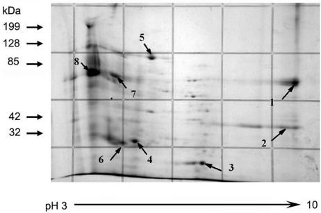
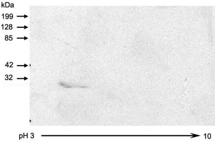
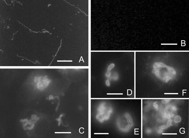
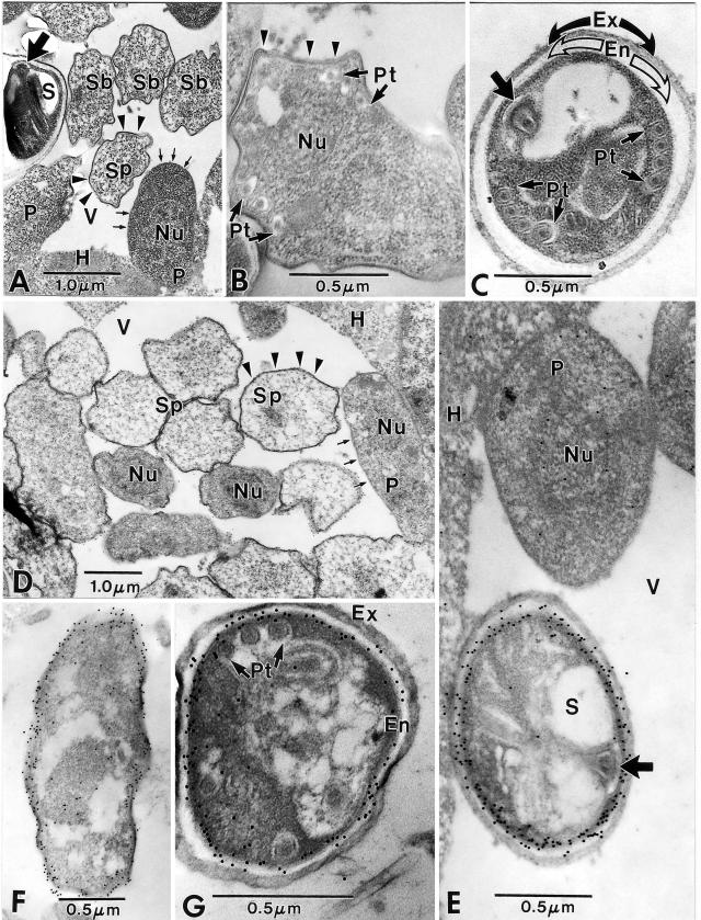
Microsporidia form environmentally resistant spores that are critical for their host-to-host transmission and persistence in the environment. The spore walls of these organisms are composed of two layers, the exospore and the endospore. Two spore wall proteins (SWP1 and SWP2) have been previously identified in members of the Encephalitozoonidae family. These proteins localize to the exospore. The endospore is known to contain chitin, and a putative glycosylphosphatidylinositol (GPI)-anchored chitin deacetylase has been localized to the plasmalemma-endospore interface. Using proteomic techniques, we have identified a new spore wall protein (SWP3) that is located in the endospore. The gene for this protein is located on chromosome 1 and corresponds to the open reading frame ECU01_1270. SWP3 is predicted to have a signal peptide and to be GPI anchored. Consistent with these modifications, two-dimensional electrophoresis demonstrated that SWP3 has an acidic pI and a molecular mass of <20 kDa. By immunoelectron microscopy, this protein was found on the cell surface during sporogony and in the endospore in mature spores. SWP3 has several potential O-glycosylation sites, and it is possible that it is a mannosylated protein like the major polar tube protein (PTP1).


Figures

FIG. 1.
FIG. 1.
Two-dimensional electrophoresis of DTT-solubilized polar tube protein preparation. The DTT-solubilized polar tube preparation was subjected to IEF using the Bio-Rad IEF system followed by polyacrylamide gel electrophoresis. The gel was stained with Coomassie blue. The arrows point to eight spots (Ec1 through Ec8) selected for mass spectrometry (mass mapping). Ec8 in the immunoblot corresponds to PTP1. Ec6 corresponds to ECU01_1270. In addition to the eight selected spots, many other protein spots can be seen in this DTT-solubilized polar tube preparation.
FIG. 2.
FIG. 2.
Amino acid sequence of ECU01_1270 (i.e., SWP3). The signal sequence (first 17 amino acids) is indicated in bold. The predicted omega cleavage site (serine 193) is shaded, and the GPI anchor signal sequence is underlined. There is a KS-rich region in the C-terminal region of this protein. The GenBank accession number for this protein is CAD25000.
FIG. 3.
FIG. 3.
Purification of recombinant ECU01_1270. (A) Coomassie blue-stained SDS-PAGE gel containing recombinant ECU01_1270 protein after elution from a glutathione-Sepharose 4B column. Bands for the full-length recombinant protein (∼51 kDa) and free GST (∼29 kDa) are evident. The presence of free GST may be due to the use of the full-length reading frame of ECU01_1270, including the N-terminal signal sequence. (B) Immunoblot of SDS-PAGE gel of recombinant ECU01_1270 protein, using a monoclonal antibody against the GST label. The full-length (∼51 kDa) recombinant protein and free GST (∼29 kDa) reacted with the anti-GST monoclonal antibody.
FIG. 4.
FIG. 4.
Immunoblot of 2D-IEF-PAGE of DTT-solubilized protein preparation. The mouse polyclonal antiserum to recombinant ECU01_1270 localizes to the protein spot Ec6 identified previously by 2D-IEF-PAGE (see Fig. 1).
FIG. 5.
FIG. 5.
Immunofluorescence microscopy of Encephalitozoon cuniculi in tissue culture. (A) Anti-EhPTP1 serum (1:100) demonstrating staining of E. cuniculi polar tubes. Bar, 10 μm. (B) There is no reactivity of anti-GST serum (1:50) with this organism. Bar, 10 μm. (C) Photomicrograph (×400) of the reactivity of anti-recombinant ECU01_1270 serum with E. cuniculi. This serum reacts with the spore wall and also stains some elongated proliferating forms at the edge of the vacuole (consistent with staining sporont membranes). Bar, 10 μm. (D) Photomicrograph (×1,000) of the reactivity of anti-recombinant ECU01_1270 serum with mature spores, demonstrating staining of the spore wall. (E to G) Photomicrograph (×1,000) of RK13 cells containing proliferating stages of E. cuniculi staining with anti-recombinant ECU01_1270 serum. Bars, 5 μm.
FIG.6.
FIG.6.
Immunoelectron microscopy of Encephalitozoon cuniculi using anti-ECU01_1270. (A to C) Control sections of Encephalitozoon cuniculi-infected rabbit kidney cells. (A) Low-magnification image of a portion of host cell cytoplasm (H) containing a parasitophorous vacuole (V) with proliferative parasite cells (P) abutted to the edge of the host-parasite interface. These cells have a simple cytoplasm, are uninucleate (Nu), and have a typical “thin” plasmalemmal surface (short arrows). The sporonts (Sp) and sporoblasts (Sb) have a “thick” dense surface coat (arrowheads) and a more complex and dense cytoplasm. The spore (S) contains a polar tube, and the anterior anchoring disk (broad arrow) is present. (B) Sporoblast containing portions of the developing polar tube (Pt), a single nucleus (Nu), and a “thick” surface coat (arrowheads). Note the irregular shape of the cell and the more complex cytoplasm. (C) Spore demonstrating groups of four cross sections of the polar tube (Pt) and its anterior anchoring disk (broad arrow). The sporoplasm is enclosed by a wide electron-lucent endospore (En), which is in turn covered by a dense, thick exospore (Ex). Note the absence of gold particles in all three images (A, B, and C). (D to G) Sections of Encephalitozoon cuniculi-infected rabbit kidney cells incubated with anti- ECU01_1270. (D) Portion of a parasitophorous vacuole (V) in the host cell cytoplasm (H) with proliferative parasite cells (P) abutted to the edge of the host-parasite interface. These cells are uninucleate (Nu) and have a typical “thin” plasmalemmal surface (short arrows). The sporonts (Sp) have a “thick” dense surface coat (arrowheads) and a more complex and dense cytoplasm. Colloidal gold particles (12 nm) are dispersed throughout the cytoplasm of the proliferative cells, while the sporogonic cells tend to have more gold localized near their plasmalemmal surfaces. There are a few gold particles at the host cell-parasite interface, but the vacuolar space (V) is free of gold. (E) High-magnification image of a portion of the host cell cytoplasm (H) and a parasitophorous vacuole (V) containing a proliferative parasite cell (P) abutted to the edge of the host-parasite interface. This uninucleate (Nu) cell has several gold particles in its cytoplasm and in the vicinity of the nucleus. The spore (S) with an anchoring disk (broad arrow) next to the proliferative cell has extensive quantities of gold distributed almost exclusively along the periphery of the plasmalemma-endospore interface. The host cell cytoplasm has a few scattered gold particles at the interface with the vacuolar space. (F) Sporont with extensive gold particles distributed along the periphery of the cell and abutting the plasmalemma and the overlying “thick” surface coat that is present during the transition to sporogony. The cytoplasm still has some diffuse gold particles in it. (G) Typical late sporoblast/early spore containing several cross sections of the polar tube and a dense sporoplasm enclosed by a well-defined plasmalemma abutted to a wide electron-lucent endospore (En) that is in turn covered by the dense exospore (Ex). A few gold particles are present in the sporoplasm, but the vast majority of them are distributed at the endospore-plasmalemma interface.

Similar articles

Cited by

References

    1. Beckers, P. J., G. J. Derks, T. Gool, F. J. Rietveld, and R. W. Sauerwein. 1996. Encephalocytozoon intestinalis-specific monoclonal antibodies for laboratory diagnosis of microsporidiosis. J. Clin. Microbiol. 34:282-285. - PMC - PubMed
    1. Biderre, C., A. Mathis, P. Deplazes, R. Weber, G. Metenier, and C. P. Vivares. 1999. Molecular karyotype diversity in the microsporidian Encephalitozoon cuniculi. Parasitology 118:439-445. - PubMed
    1. Bigliardi, E., M. G. Selmi, P. Lupetti, S. Corona, S. Gatti, M. Scaglia, and L. Sacchi. 1996. Microsporidian spore wall: ultrastructural findings on Encephalitozoon hellem exospore. J. Eukaryot. Microbiol. 43:181-186. - PubMed
    1. Bohne, W., D. J. Ferguson, K. Kohler, and U. Gross. 2000. Developmental expression of a tandemly repeated, glycine- and serine-rich spore wall protein in the microsporidian pathogen Encephalitozoon cuniculi. Infect. Immun. 68:2268-2275. - PMC - PubMed
    1. Brosson, D., L. Kuhn, G. Prensier, C. P. Vivares, and C. Texier. 2005. The putative chitin deacetylase of Encephalitozoon cuniculi: a surface protein implicated in microsporidian spore-wall formation. FEMS Microbiol. Lett. 247:81-90. - PubMed

Publication types

MeSH terms