. 2006 Jan;74(1):382-9.
doi: 10.1128/IAI.74.1.382-389.2006.

Glyceraldehyde-3-phosphate dehydrogenase of Paracoccidioides brasiliensis is a cell surface protein involved in fungal adhesion to extracellular matrix proteins and interaction with cells

Affiliations

Glyceraldehyde-3-phosphate dehydrogenase of Paracoccidioides brasiliensis is a cell surface protein involved in fungal adhesion to extracellular matrix proteins and interaction with cells

Mônica Santiago Barbosa et al. Infect Immun. 2006 Jan.

Abstract

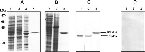
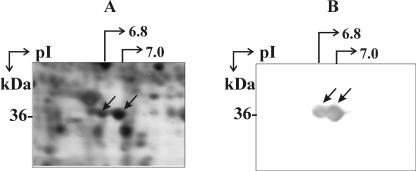
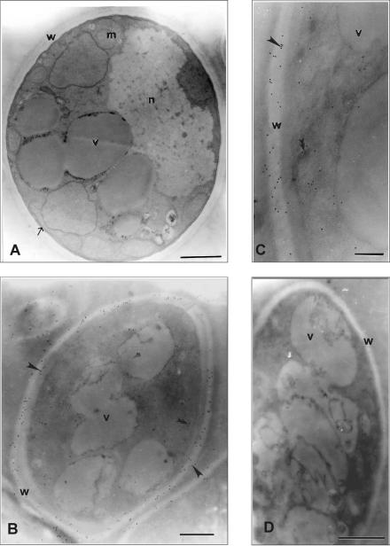
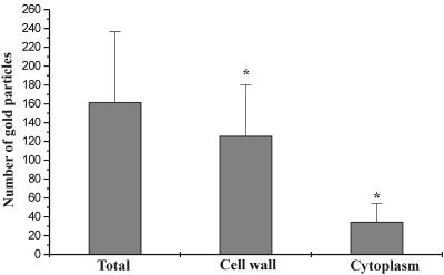
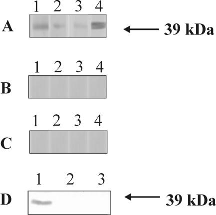
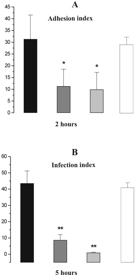
The pathogenic fungus Paracoccidioides brasiliensis causes paracoccidioidomycosis, a pulmonary mycosis acquired by inhalation of fungal airborne propagules, which may disseminate to several organs and tissues, leading to a severe form of the disease. Adhesion to and invasion of host cells are essential steps involved in the infection and dissemination of pathogens. Furthermore, pathogens use their surface molecules to bind to host extracellular matrix components to establish infection. Here, we report the characterization of the glyceraldehyde-3-phosphate dehydrogenase (GAPDH) of P. brasiliensis as an adhesin, which can be related to fungus adhesion and invasion. The P. brasiliensis GAPDH was overexpressed in Escherichia coli, and polyclonal antibody against this protein was obtained. By immunoelectron microscopy and Western blot analysis, GAPDH was detected in the cytoplasm and the cell wall of the yeast phase of P. brasiliensis. The recombinant GAPDH was found to bind to fibronectin, laminin, and type I collagen in ligand far-Western blot assays. Of special note, the treatment of P. brasiliensis yeast cells with anti-GAPDH polyclonal antibody and the incubation of pneumocytes with the recombinant protein promoted inhibition of adherence and internalization of P. brasiliensis to those in vitro-cultured cells. These observations indicate that the cell wall-associated form of the GAPDH in P. brasiliensis could be involved in mediating binding of fungal cells to fibronectin, type I collagen, and laminin, thus contributing to the adhesion of the microorganism to host tissues and to the dissemination of infection.


Figures

FIG. 1.
FIG. 1.
Expression and purification of recombinant GAPDH and generation of rabbit polyclonal antibody. (A) SDS-PAGE analysis of P. brasiliensis recombinant GAPDH. E. coli cells harboring the TOPO-pET-100 GAPDH plasmid were grown at 37°C to an A600 of 0.6 and harvested before (lane 2) and after (lane 3) a 2-h incubation with 0.8 mM IPTG. The cells were lysed by extensive sonication. Lane 1, control E. coli cells; lane 4, affinity-isolated GAPDH. SDS-12% PAGE was carried out, and the proteins were stained by Coomassie blue R-250. (B) Electrophoretic analysis of P. brasiliensis proteins and recombinant GAPDH. The protein extracts were fractionated by one-dimensional gel electrophoresis and stained by Coomassie blue. Lane 1, protein extracts from yeast cells (30 μg); lane 2, protein extracts from mycelium (30 μg); lane 3, recombinant GAPDH (2.0 μg). (C and D) Western blot analysis of native and recombinant GAPDH. The same samples as for panel B were run in parallel, blotted onto a nitrocellulose membrane, and detected by using (C) rabbit polyclonal anti-recombinant GAPDH antibody or (D) rabbit preimmune serum. After reactions with the anti-rabbit IgG alkaline phosphatase-coupled antibody (diluted 1:1,000), the reactions were developed with BCIP-NBT. Molecular size markers are indicated.
FIG. 2.
FIG. 2.
Two-dimensional gel electrophoresis of P. brasiliensis cell extracts. Cell extracts from yeast cells of P. brasiliensis were obtained, and 50 μg of total proteins was loaded on two-dimensional gels. (A) SDS-PAGE gel stained with silver. (B) Reactivity of the GAPDH isoforms analyzed by Western blotting with the polyclonal antibody produced to the recombinant protein. Arrows point to the two characterized isoforms of GAPDH. Numbers to the left of both figures refer to the molecular mass of the characterized GAPDH. At the top are indicated the isoelectric points of both protein isoforms.
FIG. 3.
FIG. 3.
Immunoelectron microscopy detection of GAPDH in P. brasiliensis yeast cells. (A) Transmission electron microscopy of P. brasiliensis yeast cells, showing the nucleus (n), intracytoplasmic vacuoles (v), and mitochondria (m). The plasma membrane (arrow) and cell wall (w) are also shown. (B and C) Gold particles (arrowheads) are observed at the fungus cell wall (w) and in the cytoplasm (double arrowheads). (D) Negative control exposed to the rabbit preimmune serum. Bars, 1 μm (A), 0.5 μm (B and D), and 0.2 μm (C).
FIG. 4.
FIG. 4.
Mean gold labeling of yeast cells of P. brasiliensis. Results, which are representative of five independent preparations, are expressed as the mean gold particles in total cells, cell wall, and cytoplasm. *, P of <0.05 in a comparison of gold labeling in the cell wall and cytoplasm. Vertical bars indicate standard deviations.
FIG. 5.
FIG. 5.
Binding of P. brasiliensis recombinant GAPDH to extracellular matrix components and to pneumocytes in culture. (A) Recombinant GAPDH (0.5 μg) was subjected to SDS-PAGE and electroblotted. Membranes were reacted with laminin (lane 1), fibronectin (lane 2), and type I collagen (lane 3) and subsequently incubated with rabbit IgG antilaminin, antifibronectin, and anti-type I collagen antibodies, respectively. Use of peroxidase-conjugated anti-rabbit IgG revealed the reactions. The positive control was obtained by incubating the recombinant protein with anti-GAPDH polyclonal antibody (lane 4). (B) Recombinant GAPDH (0.5 μg) was subjected to SDS-PAGE and electroblotted. The membranes were reacted with rabbit IgG antilaminin, antifibronectin, and anti-type I collagen antibodies (lanes 1, 2, and 3, respectively). The recombinant protein was incubated just with the secondary antibody, peroxidase-labeled goat anti-rabbit IgG (lane 4). (C) BSA (0.5 μg) was subjected to SDS-PAGE and electroblotted. The membranes were reacted with laminin (lane 1), fibronectin (lane 2), and type I collagen (lane 3) and subsequently incubated with rabbit IgG antilaminin, antifibronectin, and anti-collagen type I antibodies, respectively. Use of peroxidase-conjugated anti-rabbit IgG revealed the reaction. BSA was incubated with anti-GAPDH polyclonal antibody, and the reaction was revealed by using anti-rabbit IgG alkaline phosphatase-coupled antibody (lane 4). (D) Cultured type II pneumocytes were incubated with 50 μg of recombinant GAPDH for 5 h at 37°C. After being washed with PBS to remove the unbound protein, the cells were lysed and the supernatant was fractionated by SDS-PAGE. After transference to membranes, immunodetection was performed by incubation with rabbit anti-GAPDH polyclonal antibody. After incubation with alkaline phosphatase-coupled anti-rabbit IgG, the reaction was developed with BCIP-NBT. Lane 1, supernatant of pneumocytes incubated with recombinant GAPDH; lane 2, pneumocytes not incubated with recombinant GAPDH; lane 3, recombinant protein added to the cell culture flask.
FIG. 6.
FIG. 6.
Interaction of P. brasiliensis yeast forms with pneumocytes. Yeast cells were pretreated for 1 h with anti-GAPDH polyclonal antibody (diluted 1:100). In addition, A549 cells were pretreated for 1 h with 25 μg/ml of recombinant GAPDH. As a control, pneumocytes were pretreated for 1 h with 25 μg/ml of BSA. (A) Adhesion of P. brasiliensis to pneumocytes was analyzed 2 h after the treatments. (B) Infection (adhesion plus internalization) of P. brasiliensis to pneumocytes was analyzed 5 h after the treatments. Black bars, control; dark gray bars, P. brasiliensis cells treated with anti-GAPDH polyclonal antibody; light gray bars, pneumocytes treated with recombinant GAPDH; white bars, pneumocytes treated with BSA. The adhesion and infection index values represent the means ± standard deviations of three independent experiments. One asterisk denotes values statistically different from the control (P < 0.05), and two asterisks denote significance at a P value of <0.0001. Vertical bars indicate standard deviations.

References

    1. André, B. C., J. D. Lopes, M. F. Franco, C. A. Vaz, and V. L. Calich. 2004. Binding of laminin to Paracoccidioides brasiliensis induces a less severe pulmonary paracoccidioidomycosis caused by virulent and low-virulence isolates. Microbes Infect. 6:549-558. - PubMed
    1. Andreotti, P. F., J. L. M. Silva, A. M. Bailão, C. M. A. Soares, G. Benard, C. P. Soares, and M. J. S. Mendes-Giannini. 2005. Isolation and partial characterization of a 30 kDa adhesin from Paracoccidioides brasiliensis. Microbes Infect. 7:875-881. - PubMed
    1. Barbosa, M. S., D. A. Cunha-Passos, M. S. S. Felipe, R. S. A. Jesuíno, M. Pereira, and C. M. A. Soares. 2004. The glyceraldehyde-3-phosphate dehydrogenase is differentially regulated in phases of Paracoccidioides brasiliensis: molecular and phylogenetic analysis. Fungal Genet. Biol. 41:667-675. - PubMed
    1. Berryman, M. A., and R. D. Rodewald. 1990. An enhanced method for post-embedding immunocytochemical staining which preserves cell membranes. J. Histochem. Cytochem. 38:159-170. - PubMed
    1. Blotta, M. H. S. L., and Z. P. Camargo. 1993. Immunological response to cell-free antigens of Paracoccidioides brasiliensis: relationship with clinical forms of paracoccidioidomycosis. J. Clin. Microbiol. 31:671-676. - PMC - PubMed

Publication types

MeSH terms

Substances