. 2006 Jan;74(1):673-81.
doi: 10.1128/IAI.74.1.673-681.2006.

CD4+-T-cell effector functions and costimulatory requirements essential for surviving mucosal infection with Citrobacter rodentium

Affiliations

CD4+-T-cell effector functions and costimulatory requirements essential for surviving mucosal infection with Citrobacter rodentium

Lynn Bry et al. Infect Immun. 2006 Jan.

Abstract

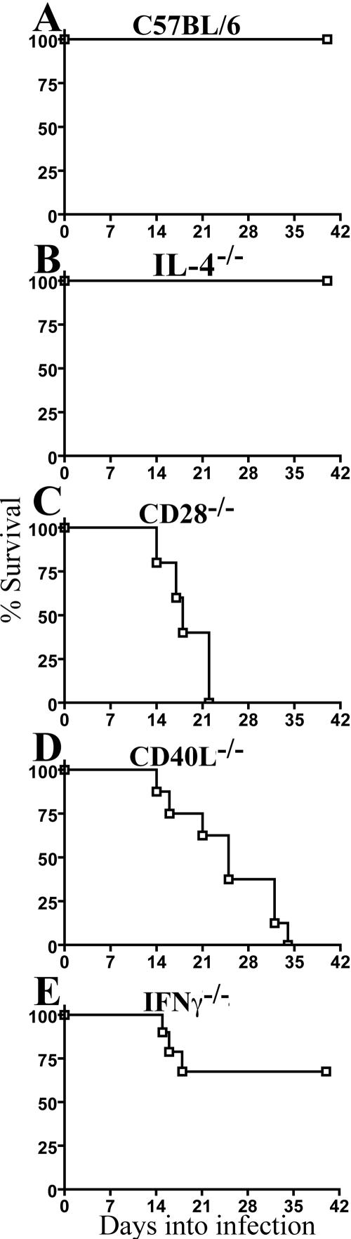
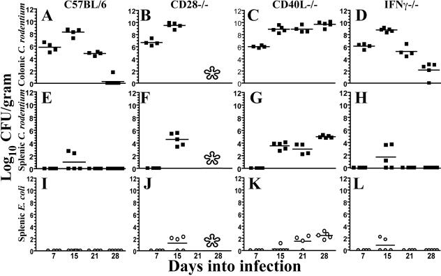
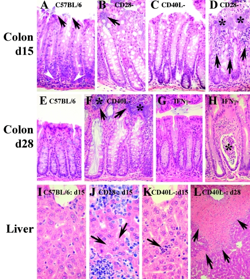
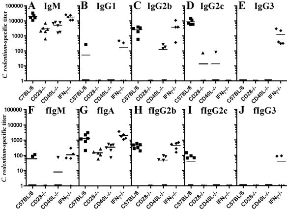
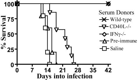
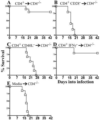
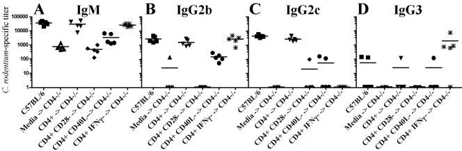
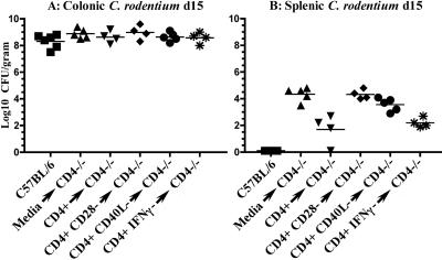
Citrobacter rodentium causes an attaching and effacing infection of the mouse colon. Surprisingly, protective adaptive immunity against this mucosal pathogen requires a systemic T-cell-dependent antibody response. To define CD4+ T-cell effector functions promoting this systemic defense of infected epithelial surfaces, studies were undertaken in weaning-age mice lacking costimulatory molecules CD28 or CD40L or cytokines gamma interferon (IFN-gamma) or interleukin-4 (IL-4). Adoptive transfer of CD4+ T cells from wild-type, CD28(-/-), CD40L(-/-), or IFN-gamma(-/-) donors to CD4(-/-) recipients delineated functions of these CD4+ T-cell-expressed molecules on the outcome of infection. Wild-type and IL-4(-/-) mice successfully resolved infection, while 70% of IFN-gamma(-/-) mice survived. In contrast, all CD28(-/-) mice succumbed during acute infection. While fewer than half of CD40L(-/-) mice succumbed acutely, surviving mice failed to clear infection, resulting in progressive mucosal destruction, polymicrobial sepsis, and death 1 to 2 weeks later than in CD28(-/-) mice. Downstream of CD28-mediated effects, CD4+ T-cell-expressed CD40L proved essential for generating acute pathogen-specific immunoglobulin M (IgM) and early IgG, which reduced pathogen burdens. However, deficiency of CD4+ T-cell-expressed IFN-gamma did not adversely impact survival or development of protective antibody in adoptively transferred CD4(-/-) recipients, though it impacted Th1 antibody responses. These findings demonstrate that CD4+ T-cell-expressed CD40L promotes the rapid production of protective systemic antibody during acute infection, while deficiencies of IL-4 or of CD4+ T-cell-expressed IFN-gamma can be overcome. These findings have important implications for understanding the role of T-helper-cell responses during infections involving mucosal surfaces.


Figures

FIG. 1.
FIG. 1.
Survival among mice lacking CD28, CD40L, IL-4, or IFN-γ. Mice were orally inoculated with 5 × 10⁸ CFU of C. rodentium at day 21 of age. (A) Wild-type C57BL/6 mice (n = 12 mice). (B) IL-4−/− mice (n = 6 mice). (C) CD28−/− mice (n = 6 mice). (D) CD40L−/− mice (n = 9 mice). (E) IFN-γ−/− mice (n = 7 mice).
FIG. 2.
FIG. 2.
Pathogen burden in colon and spleen. The x axis denotes the number of days into infection. The y axis denotes log10 CFU of C. rodentium/gram of colon (A to D) or spleen (E to H) or E. coli in spleen (I to L). Lower limits of detection are 20 CFU/gram spleen and 12 CFU/gram colon. An asterisk indicates no surviving mice at those time points for analysis. Squares, CFU of C. rodentium; open circles, CFU of E. coli.
FIG. 3.
FIG. 3.
Tissue pathology in infected mouse strains. Shown are hematoxylin and eosin stains at a ×200 magnification unless otherwise noted. (A) Colon of C57BL/6 mouse at day 15 of infection showing adherent C. rodentium (arrows) and lymphocytic infiltrate near the epithelial crypts (arrowheads). The epithelium remains intact. (B) Adherent C. rodentium (arrow) in a CD28−/− mouse. (C) CD40L−/− mouse at day 15. (D) Area of disrupted epithelium in a CD28−/− mouse at 15 days into infection. Asterisks indicate microcolonies of C. rodentium in the lamina propria. Arrows point to abscesses and epithelial disruptions. (E) Colon of wild-type mouse 28 days into infection. C. rodentium cells have been cleared, and epithelial hyperplasia has largely resolved, though remnants of the crypt-associated inflammatory infiltrate remain. (F) Colon of CD40L−/− mouse at 28 days showing mucin-engorged crypts, epithelial disruptions (arrows), and microcolonies of C. rodentium contacting the mucosa (asterisk). (G) Colon from IFN-γ−/− mouse (similar to panel E). (H) Rare lamina propria microabscess in IFN-γ−/− mouse 28 days into infection with sloughed cells in the crypt lumen (asterisk). (I to L) Liver at ×400. (I) Clear hepatic sinusoid from wild-type mouse at day 15 of infection. The top of the image shows the central vein. (J) Sinusoids from CD28−/− mouse demonstrating abscess formation. Arrows point to an area of ballooning degeneration. (K) Section from CD40L−/− mouse similar to panel I, but demonstrating a small microabscess (arrow). (L) A ×100 image of liver from a CD40L−/− mouse at day 28 showing widespread geographic necrosis (arrows) extending from regions supplied by the portal circulation.
FIG. 4.
FIG. 4.
Anti-C. rodentium serum and mucosal Ig responses: the x axis denotes mouse strains, and the y axis shows relative endpoint titers. Bars indicate mean values. Squares, wild-type C57BL/6 mice; triangles, CD28−/− mice; inverted triangles, CD40L−/− mice; diamonds, IFN-γ−/− mice. (A to E) Serum antibody responses against C. rodentium at day 15 of infection. (A) IgM. (B) IgG1. (C) IgG2b. (D) IgG2c. (E) IgG3. (F to J) Mucosal antibody responses against C. rodentium at day 15. (F) Fecal IgM. (G) IgA. (H) IgG2b. (I) IgG2c. (J) IgG3. The minimum detectable titer is 50.
FIG. 5.
FIG. 5.
Transfer of acute-phase serum to CD4−/− recipients. Shown are results from CD4−/− mice receiving saline (squares; 5 mice), preimmune serum from naïve C57BL/6 mice (triangles; 5 mice), or serum from acutely infected wild-type (crosses; 5 mice), CD40L−/− (inverted triangles; 7 mice), or IFN-γ−/− mice (diamonds; 5 mice). The crosses (representing wild type) and diamonds (representing IFN-γ−/− donors) overlap.
FIG. 6.
FIG. 6.
Survival of CD4−/− mice adoptively transferred with CD4+ T cells from wild-type or CD28-, CD40L-, or IFN-γ-deficient donors. (A) Mice receiving wild-type CD4+ T cells (n = 10 mice). (B) CD4+ CD28 T cells (n = 5 mice). (C) CD4+ CD40L T cells (n = 6 mice). (D) CD4+ IFN-γ T cells (n = 5 mice). (E) CD4−/− recipients receiving media (n = 8 mice).
FIG. 7.
FIG. 7.
Acute antibody responses in adoptively transferred CD4−/− mice. Shown are pathogen-specific IgM (A), IgG2b (B), IgG2c (C), and IgG3 (D) antibody responses. The x axis indicates antibody responses in wild-type, CD4−/−, or adoptively transferred CD4−/− recipients; The y axis indicates the relative endpoint titer. The minimum detectable titer is 50. Squares, wild-type C57BL/6 mice; triangles, CD4−/− recipients receiving medium only; inverted triangles, CD4−/− recipients receiving wild-type CD4 T cells; diamonds, CD4−/− recipients receiving CD28−/− CD4+ T cells; circles, CD4−/− recipients receiving CD40L−/− CD4+ T cells; asterisks, CD4−/− recipients receiving IFN-γ−/− CD4+ T cells.
FIG. 8.
FIG. 8.
Burden of C. rodentium in adoptively transferred CD4−/− mice. Shown are colonic (A) and splenic (B) CFU of C. rodentium at 15 days of infection. The y axis denotes log10 CFU of C. rodentium per gram of tissue. The x axis indicates the mouse strain and cell populations transferred to CD4−/− mice. Squares, wild-type C57BL/6 mice; triangles, CD4−/− recipients receiving medium only; inverted triangles, CD4−/− recipients receiving wild-type CD4 T cells; diamonds, CD4−/− recipients receiving CD28−/− CD4+ T cells; circles, CD4−/− recipients receiving CD40L−/− CD4+ T cells; asterisks, CD4−/− recipients receiving IFN-γ−/− CD4+ T cells.

Similar articles

Cited by

References

    1. Bertram, E. M., A. Tafuri, A. Shahinian, V. S. Chan, L. Hunziker, M. Recher, P. S. Ohashi, T. W. Mak, and T. H. Watts. 2002. Role of ICOS versus CD28 in antiviral immunity. Eur. J. Immunol. 32:3376-3385. - PubMed
    1. Bry, L., and M. B. Brenner. 2004. Critical role of T cell-dependent serum antibody, but not the gut-associated lymphoid tissue, for surviving acute mucosal infection with Citrobacter rodentium, an attaching and effacing pathogen. J. Immunol. 172:433-441. - PubMed
    1. Clark, M. A., B. H. Hirst, and M. A. Jepson. 1998. M-cell surface β1 integrin expression and invasin-mediated targeting of Yersinia pseudotuberculosis to mouse Peyer's patch M cells. Infect. Immun. 66:1237-1243. - PMC - PubMed
    1. Cleary, J., L. C. Lai, R. K. Shaw, A. Straatman-Iwanowska, M. S. Donnenberg, G. Frankel, and S. Knutton. 2004. Enteropathogenic Escherichia coli (EPEC) adhesion to intestinal epithelial cells: role of bundle-forming pili (BFP), EspA filaments and intimin. Microbiology 150:527-538. - PubMed
    1. Deenick, E. K., J. Hasbold, and P. D. Hodgkin. 1999. Switching to IgG3, IgG2b, and IgA is division linked and independent, revealing a stochastic framework for describing differentiation. J. Immunol. 163:4707-4714. - PubMed

Publication types

MeSH terms

LinkOut - more resources