. 2006 Jan;38(1):38-46.
doi: 10.1038/ng1698. Epub 2005 Dec 20.

Homozygous L-SIGN (CLEC4M) plays a protective role in SARS coronavirus infection

Affiliations

Homozygous L-SIGN (CLEC4M) plays a protective role in SARS coronavirus infection

Vera S F Chan et al. Nat Genet. 2006 Jan.

Abstract

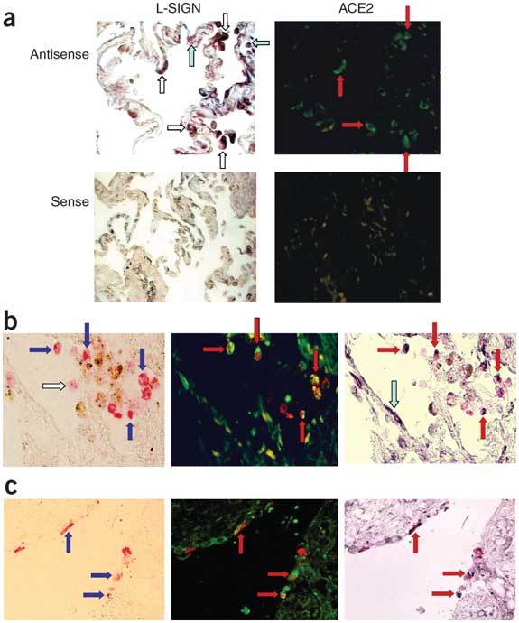
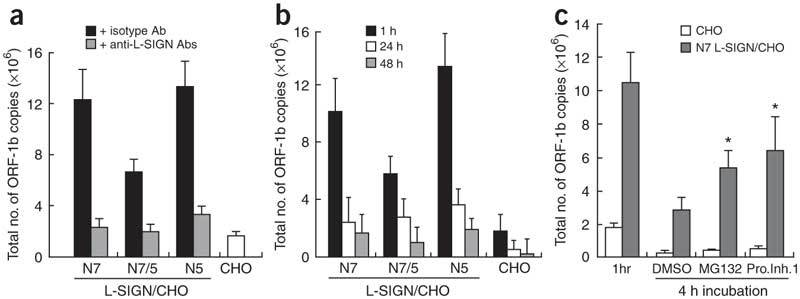
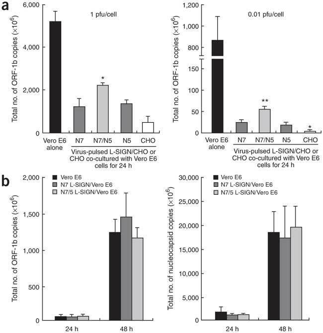
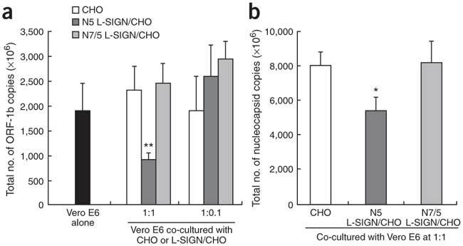
Severe acute respiratory syndrome (SARS) is caused by infection of a previously undescribed coronavirus (CoV). L-SIGN, encoded by CLEC4M (also known as CD209L), is a SARS-CoV binding receptor that has polymorphism in its extracellular neck region encoded by the tandem repeat domain in exon 4. Our genetic risk association study shows that individuals homozygous for CLEC4M tandem repeats are less susceptible to SARS infection. L-SIGN is expressed in both non-SARS and SARS-CoV-infected lung. Compared with cells heterozygous for L-SIGN, cells homozygous for L-SIGN show higher binding capacity for SARS-CoV, higher proteasome-dependent viral degradation and a lower capacity for trans infection. Thus, homozygosity for L-SIGN plays a protective role during SARS infection.


Conflict of interest statement

The authors declare no competing financial interests.

Figures

Figure 1
Figure 1. L-SIGN is expressed in non-SARS lung and also in the lung and small bowel of patients fatally infected with SARS.
(a) In situ hybridization for L-SIGN in paraffin-embedded non-SARS lung tissues was performed with an L-SIGN specific antisense RNA (cRNA) probe (dark purple, upper left panel). Fluorescence in situ hybridization (FISH) for ACE2 was also performed on the same section (shown in green, upper right panel). L-SIGNbright cells either do (white arrows, upper left panel versus red arrows, upper right panel) or do not (sky blue arrows, upper left panel) co-express ACE2. Sense controls are shown in lower panels respectively. (b) Paraffin-embedded SARS lung samples were first stained with monoclonal antibody against the SARS-CoV nucleocapsid antigen and anti-mouse universal immunoalkalinephosphatase polymer, and the color was subsequently developed with fast red substrate system (shown in red, left panel). FISH for ACE2 (shown in green, middle panel) and in situ hybridization for L-SIGN (shown in dark purple, right panel) were also performed on the same section. SARS-CoV antigen was identified in L-SIGNbrightACE2bright cells (blue arrows, left panel versus red arrows, middle panel versus red arrows, right panel). No viral antigens were detected in L-SIGNbrightACE2dim/− cells (sky blue arrows, right panel). SARS-CoV antigen was identified in L-SIGNdim/−ACE2dim/− cells (left panel, white arrows). (c) Similar staining procedures were conducted on small bowel samples from SARS patients as described in b. SARS-CoV antigen could be identified in L-SIGNbrightACE2bright cells on luminal surface of small bowel of patients fatally infected with SARS (blue arrows, left panel versus red arrows, middle panel versus red arrows, right panel).
Figure 2
Figure 2. L-SIGN is a binding receptor for SARS-CoV and mediates proteasome-dependent viral degradation.
(a) Viral ORF-1b copy number of N7, N5 and N7/5 L-SIGN transfected CHO cells (L-SIGN/CHO), and mock transfectants (CHO), pre-treated with anti-L-SIGN antibodies (clones 120526 and 120612, 10 μg/ml each) or isotype control antibody before being pulsed with 1 PFU/cell SARS-CoV and incubated for 1 h at 4 °C and washed. (b) Total viral ORF-1b copy number (cell lysates plus supernatants) of N7, N5 and N7/5 L-SIGN transfected CHO cells, and mock transfectants, pulsed with 1 PFU/cell SARS CoV for 1h, washed and subsequently incubated at 37 °C for 24 and 48h. (c) Total viral ORF-1b copy number of N7 L-SIGN transfected CHO cells, and mock transfectants, pulsed with 1 PFU/cell SARS CoV for 1 h at 37 °C, washed and subsequently incubated for 4 h in the presence or absence of 10 μM MG132 or proteasome inhibitor I (Pro. Inh. 1). *P < 0.05 in comparison to cells treated with DMSO solvent control. Data are expressed as mean ± s.d. from triplicate, and are representative of 3 experiments.
Figure 3
Figure 3. L-SIGN facilitates trans, but not cis, infection of SARS-CoV.
(a) N7, N5 and N7/5 L-SIGN/CHO and mock transfected CHO cells at 1 × 10⁴/well in 96-well plates were pulsed with 1 PFU/cell (left panel) or 0.01 PFU/cell (right panel) SARS-CoV for 1 h at 37 °C. After wash, cells were cultured with 1 × 10⁴ Vero E6 cells for 24 h and the total copy number of ORF-1b transcripts was determined. *P < 0.05 in comparison with homozygous N7 or N5 L-SIGN/CHO (left panel). *P < 0.05 and **P < 0.01 in comparison with homozygous N7 or N5 L-SIGN/CHO (right panel). (b) N7 and N7/N5 L-SIGN transfected Vero E6 cells and mock transfected Vero E6 cells were exposed to 0.01 PFU/cell SARS-CoV for 1 h at 37 °C. After wash, cells were incubated for 24 h and 48 h and the total copy number of viral ORF-1b (left panel) and nucleocapsid (right panel) transcripts were determined.
Figure 4
Figure 4. L-SIGN expressed on permissive Vero E6 cells captured/adsorbed infectious viruses released into the supernatant.
(a) N7, N7/N5 L-SIGN transfected Vero E6 cells and mock transfectant Vero E6 cells were infected with 0.01 PFU/cell SARS-CoV for 1 h at 37 °C. After wash, cells were incubated for 24 h and 48 h before cells were harvested separately from supernatant for the quantification of the viral ORF-1b copy number in cell lysates and supernatant. *P < 0.05; **P < 0.01 in comparison to heterozygous N7/5 L-SIGN/Vero E6 and mock transfected Vero E6. (b) N7 L-SIGN transfected Vero E6 cells were pre-treated with anti-L-SIGN antibodies or isotype control antibody, followed by infection with 0.01 PFU/cell SARS-CoV for 1 h at 37 °C. After wash, cells were further incubated for 48 h in the presence of anti-L-SIGN antibodies or isotype control antibody before determination of the viral ORF-1b transcripts in cell lysates and supernatant. (c) N7 L-SIGN transfected Vero E6 cells and mock transfectants were infected with 0.01 PFU/cell SARS-CoV for 1 h at 37 °C. After wash, cells were incubated for 48 h. The N7 L-SIGN/Vero E6 cells were further treated with PBS or 5 mM EGTA for 1 h before harvest for quantification of ORF-1b copy number in cell lysates and supernatant. *P < 0.05 in comparison to N7 L-SIGN/Vero treated with EGTA; **P < 0.01 in comparison to N7 L-SIGN/Vero treated with PBS. Data are expressed as mean ± s.d. of triplicates, and are representative of three experiments.
Figure 5
Figure 5. Homozygous, but not heterozygous, L-SIGN reduced the final total viral genomic and subgenomic copy number in a closed infection system.
(a) 1 × 10⁴ Vero E6 cells were cultured with 1 × 10⁴ (ratio 1:1) or 1 × 10³ (ratio 1:0.1) stable transfectants of N5 or N7/5 L-SIGN transfected CHO cells, and subsequently infected with 0.01 PFU per Vero E6 cell. Without washing, cells were further incubated for 48 h before the determination of the total viral genomic ORF-1b copy number. (b) Cells were treated as in a for the determination of the total nucleocapsid copy number. Data are expressed as mean ± s.d. from triplicate and are representative of three experiments. *P < 0.05, **P < 0.01 in comparison to heterozygous N7/5 L-SIGN/CHO.

Comment in

Similar articles

Cited by

References

    1. Peiris JS, et al. Coronavirus as a possible cause of severe acute respiratory syndrome. Lancet. 2003;361:1319–1325. doi: 10.1016/S0140-6736(03)13077-2. - DOI - PMC - PubMed
    1. Ksiazek TG, et al. A novel coronavirus associated with severe acute respiratory syndrome. N. Engl. J. Med. 2003;348:1953–1966. doi: 10.1056/NEJMoa030781. - DOI - PubMed
    1. Drosten C, et al. Identification of a novel coronavirus in patients with severe acute respiratory syndrome. N. Engl. J. Med. 2003;348:1967–1976. doi: 10.1056/NEJMoa030747. - DOI - PubMed
    1. Bonavia A, Zelus BD, Wentworth DE, Talbot PJ, Holmes KV. Identification of a receptor-binding domain of the spike glycoprotein of human coronavirus HCoV-229E. J. Virol. 2003;77:2530–2538. doi: 10.1128/JVI.77.4.2530-2538.2003. - DOI - PMC - PubMed
    1. Breslin JJ, et al. Human coronavirus 229E: receptor binding domain and neutralization by soluble receptor at 37 degrees C. J. Virol. 2003;77:4435–4438. doi: 10.1128/JVI.77.7.4435-4438.2003. - DOI - PMC - PubMed

Publication types

MeSH terms

Associated data