

Blood flow in the common carotid artery in term and preterm infants: reproducibility and relation to cardiac output
- PMID: 16371390
- PMCID: PMC2672646
- DOI: 10.1136/adc.2004.058172
Blood flow in the common carotid artery in term and preterm infants: reproducibility and relation to cardiac output
Abstract
Aim: To assess the reproducibility of, and determine normative data for, flow volume measurements from the right common carotid artery (CCA) and its relation to left ventricular output (LVO) in stable term and preterm babies using Doppler ultrasound.
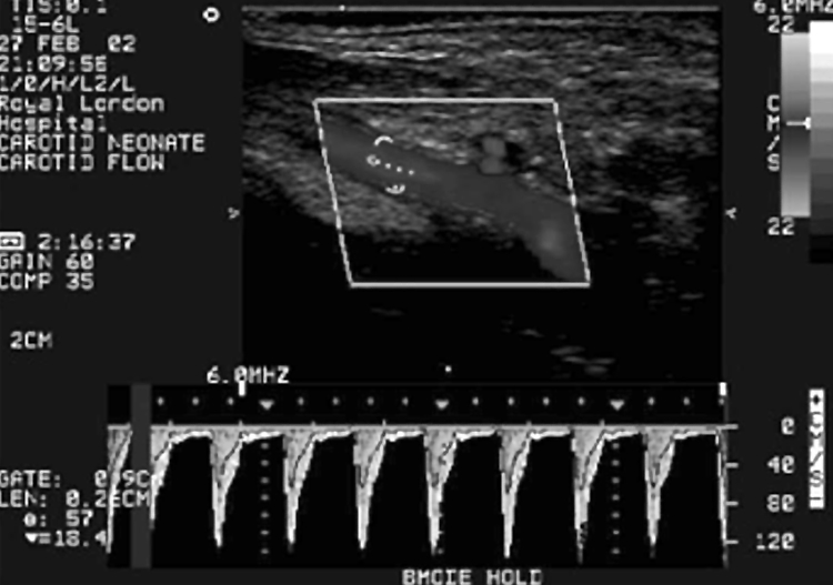
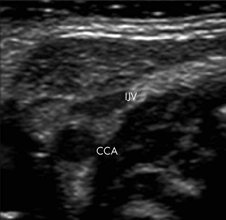
Methods: Right CCA flow volume was measured using a near focus, high frequency transducer by obtaining intensity weighted mean velocity and right CCA diameter. LVO was determined using standard Doppler techniques. Reproducibility studies were performed on 30 newborn infants by two observers. Normative data were obtained from 40 spontaneously breathing preterm babies and 21 term babies.
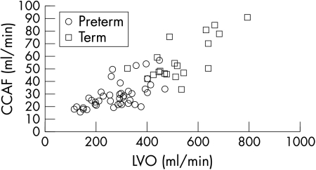
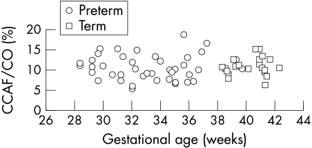
Results: The intraobserver coefficient of variation for CCA flow measurements was 10.5% for observer 1 and 15.4% for observer 2, whereas the interobserver coefficient of variation was 16.4%. In term and preterm infants, right CCA flow was about 20 ml/kg/min, accounting for 11% of cardiac output. Among the preterm infants, there was a positive correlation of right CCA flow with gestation (r = 0.61, p<0.001), weight (r = 0.64, p<0.001), and LVO (r = 0.59, p<0.001). Right CCA diameter also increased with weight (r = 0.63, p<0.001) and gestation (r = 0.58, p<0.001). The proportion of LVO distributed to the right CCA did not increase with gestation, nor did the right CCA flow per kg body weight.
Conclusions: It is possible to perform reproducible measurements of flow volume in the CCA of newborn infants. In stable, spontaneously breathing babies, both cardiac output and carotid flow increased with gestation and body weight. The proportion of cardiac output distributed to the right CCA remained relatively constant across gestation.
Conflict of interest statement
Competing interests: none declared
Similar articles
-
[Clinical study of neonatal cardiac output measurement methods].Zhonghua Er Ke Za Zhi. 2013 Jan;51(1):58-63. Zhonghua Er Ke Za Zhi. 2013. PMID: 23527933 Chinese.
-
Central blood flow measurements in stable preterm infants after the transitional period.Arch Dis Child Fetal Neonatal Ed. 2010 Sep;95(5):F369-72. doi: 10.1136/adc.2009.169169. Epub 2010 Jun 7. Arch Dis Child Fetal Neonatal Ed. 2010. PMID: 20530103
-
Reproducibility of measurements of cardiac output in newborn infants by Doppler ultrasound.Arch Dis Child. 1990 Jan;65(1 Spec No):15-9. doi: 10.1136/adc.65.1_spec_no.15. Arch Dis Child. 1990. PMID: 2407197 Free PMC article.
-
A pilot study of the relationship between Doppler-estimated carotid and brachial artery flow and cardiac index.Anaesthesia. 2015 Oct;70(10):1140-7. doi: 10.1111/anae.13069. Epub 2015 May 25. Anaesthesia. 2015. PMID: 26010229
-
Supporting preterm cardiovascular function.Clin Exp Pharmacol Physiol. 2019 Mar;46(3):274-279. doi: 10.1111/1440-1681.13044. Epub 2019 Feb 10. Clin Exp Pharmacol Physiol. 2019. PMID: 30347457 Review.
Cited by
-
A realistic flow phantom model of the carotid artery in preterm infants for training and research.Ultrasound. 2020 Aug;28(3):145-154. doi: 10.1177/1742271X20902189. Epub 2020 Feb 3. Ultrasound. 2020. PMID: 32831887 Free PMC article.
-
Investigation of EEG Activity Compared with Mean Arterial Blood Pressure in Extremely Preterm Infants.Front Neurol. 2018 Feb 26;9:87. doi: 10.3389/fneur.2018.00087. eCollection 2018. Front Neurol. 2018. PMID: 29535674 Free PMC article.
-
Distinct Cellular Profiles of Hif1a and Vegf mRNA Localization in Microglia, Astrocytes and Neurons during a Period of Vascular Maturation in the Auditory Brainstem of Neonate Rats.Brain Sci. 2021 Jul 18;11(7):944. doi: 10.3390/brainsci11070944. Brain Sci. 2021. PMID: 34356178 Free PMC article.
-
A Preterm Physiologically Based Pharmacokinetic Model. Part I: Physiological Parameters and Model Building.Clin Pharmacokinet. 2020 Apr;59(4):485-500. doi: 10.1007/s40262-019-00825-6. Clin Pharmacokinet. 2020. PMID: 31583613
-
Transmission in near-infrared optical windows for deep brain imaging.J Biophotonics. 2016 Jan;9(1-2):38-43. doi: 10.1002/jbio.201500192. Epub 2015 Nov 10. J Biophotonics. 2016. PMID: 26556561 Free PMC article.
References
-
- Pape K E, Wigglesworth J S. Haemorrhage, ischaemia and the perinatal brain. Clinics in developmental medicine. London: Spastics International Medical Publications, 19791–196.
-
- Kempley S T, Gamsu H R. Arterial blood pressure and blood flow velocity in major cerebral and visceral arteries. I. Interindividual differences. Early Hum Dev 199334227–232. - PubMed
MeSH terms
LinkOut - more resources
Full Text Sources
Medical