Comparative Study
. 2006 Jan 3;103(1):153-8.
doi: 10.1073/pnas.0509784103. Epub 2005 Dec 21.

Regulation of dendritic cell maturation and function by Bruton's tyrosine kinase via IL-10 and Stat3

Affiliations
Comparative Study

Regulation of dendritic cell maturation and function by Bruton's tyrosine kinase via IL-10 and Stat3

Yuko Kawakami et al. Proc Natl Acad Sci U S A. .

Abstract

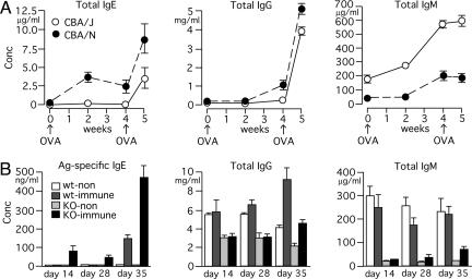
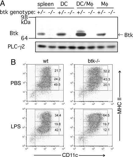
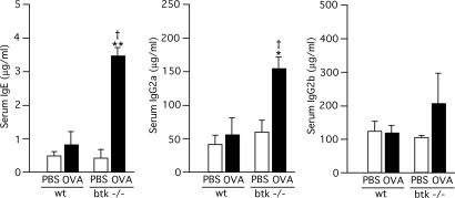
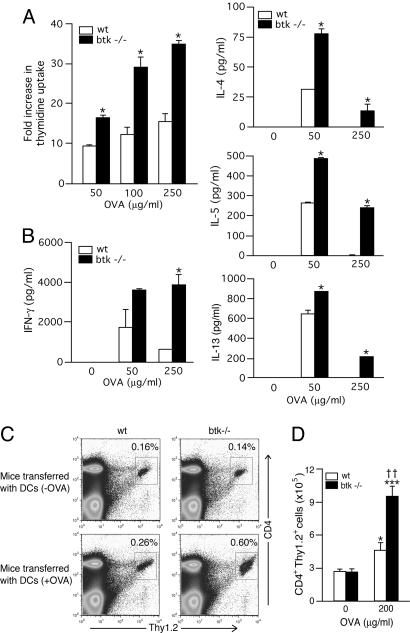
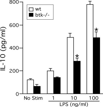
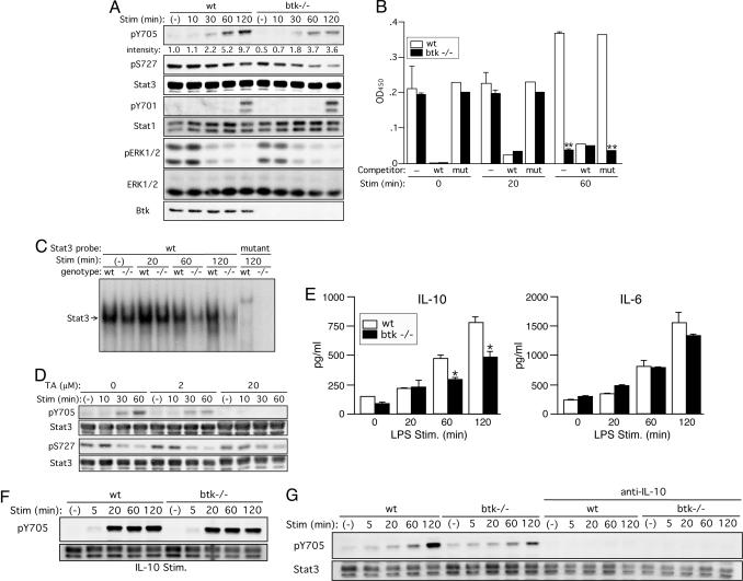
Btk plays crucial roles in the differentiation and activation of B and myeloid cells. Despite drastic reductions of other Ig isotypes, paradoxically high IgE responses have been known in btk mutant mice. Here we show that btk(-/-) dendritic cells exhibit a more mature phenotype and a stronger in vitro and in vivo T cell-stimulatory ability than wild-type cells. Increased IgE responses were induced by adoptive transfer of btk(-/-) dendritic cells into mice. Consistent with the stronger T cell-stimulatory ability of btk(-/-) dendritic cells, btk(-/-) mice exhibited enhanced inflammation in Th2-driven asthma and Th1-driven contact sensitivity experiments. These negative regulatory functions of Btk in dendritic cells appear to be mediated mainly through autocrine secretion of IL-10 and subsequent activation of Stat3.


Figures

Fig. 1.
Fig. 1.
High-serum IgE responses in btk mutant mice. (A) CBA/J(WT) and CBA/N(xid) mice were immunized with i.p. injection of 1 μg of OVA mixed with 1 mg of alum at week 0 and week 4. Sera were collected at the indicated times, and immunoglobulins were measured. Each value represents the mean ± SEM of 3–5 mice. Similar results are reproduced in another experiment. (B) WT and btk–/– mice were left unimmunized (n = 3 each) or immunized (n = 6 each) by i.p. injection of 10 μg of DNP-Asc mixed with 1 mg of alum on day 0 and day 28. Sera were collected on days 14, 28, and 35. Each value represents the mean ± SEM. Similar results are reproduced in another experiment.
Fig. 2.
Fig. 2.
Btk expression and surface expression of MHC II in WT and btk–/– BMDCs. (A) Bone marrow cells were incubated in recombinant mouse GM-CSF-containing medium for 7 days. More than 95%-pure CD11c+ BMDCs (DC) were used. Adherent cells (Mφ) after 7 days in culture and a mixed population of adherent and nonadherent cells (DC/Mφ) after 3 days in culture were also analyzed together with another control, unfractionated spleen cells (spleen). Cell lysates were analyzed by immunoblotting. The 64- to 98-kDa portion of the blot was probed with anti-Btk antibody, and the higher Mr portion with anti-PLC-γ2 antibody. (B) BMDCs were incubated with PBS or 10 ng/ml LPS for 18 h. The cells were stained with FITC-conjugated anti-CD11c and PE-conjugated anti-I-A/I-E (M5/114.15.2) mAbs before flow cytometric analysis. Percentages of cell populations in each box are shown. The MHC phenotypes shown are representative of at least 48 independent BMDC cultures.
Fig. 3.
Fig. 3.
Increased production of IgE and IgG2a in mice adoptively transferred with Btk-deficient dendritic cells. WT and btk–/– BMDCs were incubated with 100 μg/ml OVA or PBS for 3 h, and the BMDCs (3 × 10⁵ cells) were adoptively transferred to B6 mice. Total IgE, IgG2a, and IgG2b levels in sera were measured 7 days later. Data represents the mean ± SEM and analyzed by two-way ANOVA. *, P < 0.05, and **, P < 0.005 (vs. the PBS control of the same genotype); †, P < 0.05 (vs. the WT control). Similar results are reproduced in three independent experiments.
Fig. 4.
Fig. 4.
Increased in vitro and in vivo T cell stimulatory activity by btk–/– DCs. (A and B) OVA-specific naïve OTII T cells were stimulated with WT or btk–/– splenic CD11c+ cells in the presence of various concentrations of OVA for 3–4 days. (A) T cell proliferation was measured by incubating the cells with [3H]thymidine for the last 12 h of the 3-day cultures. Each value represents the mean ± SD. (B) Cytokines secreted into culture media for 4 days were measured by ELISA (mean ± SD). *, P < 0.05 (vs. the WT control; Student's t test). Similar results are reproduced four independent experiments. (C and D) OTII CD4+ (Thy1.2) cells were i.v. injected into Thy1.1 congenic B6 mice. Splenic DCs from WT and btk–/– mice were stimulated with LPS in the presence or absence of 200 μg/ml OVA for 16 h and i.v. injected into the mice 24 h after OTII CD4+ cell injection. The mice were killed 5 days later, and Thy1.2+ CD4+ T cells were enumerated by flow cytometry. Each value represents the mean ± SEM. *, P < 0.05, and ***, P < 0.0001 (vs. the DC [-OVA] control of the same genotype; two-way ANOVA); ††, P < 0.005 (vs. the WT control). Similar results are reproduced in two more independent experiments.
Fig. 5.
Fig. 5.
Reduced IL-10 production in btk–/– DCs. BMDCs were incubated with PBS or 1–100 ng/ml LPS for 24 h. IL-10 secreted into culture media was measured by ELISA. No detectable IL-4 was produced (data not shown). *, P < 0.05 (vs. the WT control; Student's t test). Results (mean ± SEM) shown are representative of five independent experiments.
Fig. 6.
Fig. 6.
Btk positively regulates Stat3 activity mainly through autocrine secretion of IL-10. (A) WT and btk–/– BMDCs were left unstimulated or stimulated with 1 μg/ml LPS for the indicated periods. The cells were lysed and analyzed by immunoblotting with anti-phospho-Stat3(Tyr-705), anti-phosho-Stat3(Ser-727), anti-phospho-Stat1(Tyr-701), anti-phospho-MAPK(Thr-202/Tyr-204), or anti-Btk. The blots were reprobed with anti-Stat3, Stat1, and anti-ERK to check for their expression. Results shown are representative of seven (Stat3) or three (others) experiments. (B) DNA-binding activities of Stat3 in BMDCs stimulated by LPS for the indicated periods were measured by ELISA-based binding assays. Specificity of Stat3 binding to the plate-bound Stat3 consensus oligonucleotide was confirmed by strong competition by free WT, but not mutant, oligonucleotide. **, P < 0.005 (vs. the WT cells; Student's t test). Similar results (mean ± SD) are reproduced in three independent experiments. (C) EMSAs were also performed by using 32P-labeled WT or mutant oligonucleotide as a probe. Position of Stat3/DNA complexes is indicated. Similar results are reproduced in three independent experiments. (D) WT BMDCs were preincubated with the indicated concentrations of terreic acid (TA) for 30 min before stimulation with LPS for the indicated periods. The cells were analyzed by immunoblotting with anti-phospho-Stat3(Tyr-705) or anti-phospho-Stat3(Ser-727). The blots were reprobed with anti-Stat3. The results shown were reproduced in two additional experiments. (E) Wt and btk–/– BMDCs were left unstimulated or stimulated with 100 ng/ml LPS for the indicated periods. Amounts of IL-10 and IL-6 secreted into medium were measured by ELISA. *, P < 0.05 (vs. the WT cells; Student's t test). Similar results (mean ± SEM) were reproduced in another experiment. (F) WT and btk–/– BMDCs were left unstimulated or stimulated with 20 ng/ml mouse recombinant IL-10 for the indicated periods. (G) WT and btk–/– BMDCs, pretreated with a neutralizing anti-IL-10 mAb for 60 min, were left unstimulated or stimulated with 1 μg/ml LPS for the indicated periods. Cell lysates were analyzed by immunoblotting for Tyr-705 phosphorylation followed by reprobing with anti-Stat3. Results shown in F and G are representative of two independent experiments.

References

    1. Banchereau, J. & Steinman, R. M. (1998) Nature 392, 245–252. - PubMed
    1. Pulendran, B., Smith, J. L., Caspary, G., Brasel, K., Pettit, D., Maraskovsky, E. & Maliszewski, C. R. (1999) Proc. Natl. Acad. Sci. USA 96, 1036–1041. - PMC - PubMed
    1. Maldonado-Lopez, R., De Smedt, T., Michel, P., Godfroid, J., Pajak, B., Heirman, C., Thielemans, K., Leo, O., Urbain, J. & Moser, M. (1999) J. Exp. Med. 189, 587–592. - PMC - PubMed
    1. Fruman, D. A., Satterthwaite, A. B. & Witte, O. N. (2000) Immunity 13, 1–3. - PubMed
    1. Smith, C. I., Baskin, B., Humire-Greiff, P., Zhou, J. N., Olsson, P. G., Maniar, H. S., Kjellen, P., Lambris, J. D., Christensson, B., Hammarstrom, L., et al. (1994) J. Immunol. 152, 557–565. - PubMed

Publication types

MeSH terms

LinkOut - more resources