Comparative Study
. 2006 Jan 3;103(1):69-74.
doi: 10.1073/pnas.0504909102. Epub 2005 Dec 21.

Intraprotein transfer of the quinone analogue inhibitor 2,5-dibromo-3-methyl-6-isopropyl-p-benzoquinone in the cytochrome b6f complex

Affiliations
Comparative Study

Intraprotein transfer of the quinone analogue inhibitor 2,5-dibromo-3-methyl-6-isopropyl-p-benzoquinone in the cytochrome b6f complex

Jiusheng Yan et al. Proc Natl Acad Sci U S A. .

Abstract

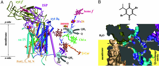
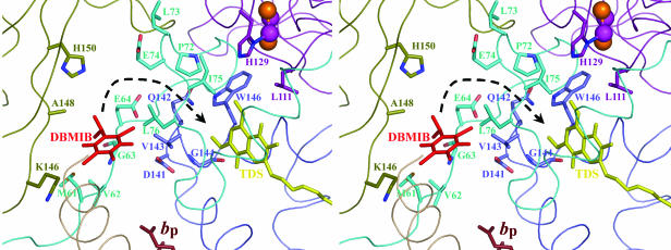
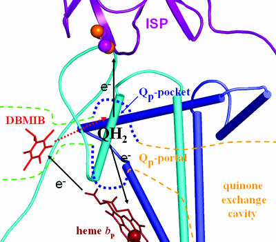
Details are presented of the structural analysis of the cytochrome b(6)f complex from the thermophilic cyanobacterium, Mastigocladus laminosus, in the presence of the electrochemically positive (p)-side quinone analogue inhibitor, 2,5-dibromo-3-methyl-6-isopropylbenzoquinone (DBMIB). One DBMIB binding site was found. This site is peripheral to the quinone binding space defined by the binding sites of other p-side inhibitors previously resolved in cytochrome bc(1)/b(6)f complexes. This high-affinity site resides in a p-side interfacial niche bounded by cytochrome f, subunit IV, and cytochrome b(6), is close (8 A) to the p-side heme b, but distant (19 A) from the [2Fe-2S] cluster. No significant electron density associated with the DBMIB was found elsewhere in the structure. However, the site at which DBMIB can inhibit light-induced redox turnover is within a few A of the [2Fe-2S] cluster, as shown by the absence of inhibition in mutants of Synechococcus sp. PCC 7002 at iron sulfur protein-Leu-111 near the cluster. The ability of a minimum amount of initially oxidized DBMIB to inhibit turnover of WT complex after a second light flash implies that there is a light-activated movement of DBMIB from the distal peripheral site to an inhibitory site proximal to the [2Fe-2S] cluster. Together with the necessary passage of quinone/quinol through the small Q(p) portal in the complex, it is seen that transmembrane traffic of quinone-like molecules through the core of cytochrome bc complexes can be labyrinthine.


Figures

Fig. 1.
Fig. 1.
Structure model of the cytochrome b6f complex with bound DBMIB. (A) Model (3.8 Å) of the dimeric b6f complex from M. laminosus with bound DBMIB. To emphasize the prosthetic groups and electron transfer pathways, (Left) all polypeptide components are shown in one monomer with α-helices viewed as cylinders, whereas (Right) only DBMIB and the prosthetic groups are shown in the other monomer. The electron density of DBMIB is shown in yellow with a contour level of 6 σ. The electron transfer pathways (see text for details) in one monomer (Right) are shown schematically. 2Fe2S, iron-sulfur cluster; blue arrows, electron transfer paths; bp, heme bp; bn, heme bn; β-Car, β-carotene; Chl a, chlorophyll a; e, electron; H+, proton; Q, quinone; QH2, quinol; Qn, n-side quinone binding site (reduction site); Qp, p-side quinone binding site (oxidation site); x, heme x. For clarity, endogenous PQ molecules bound at the Qn site are not shown. (B)(Upper) Chemical structure of DBMIB and (Lower) cross-sectional view of its binding site and position relative to the Qp pocket. To show the position of the Qp pocket, a ring-in TDS molecule (yellow) from the C. reinhardtii structure is superimposed on the structure. The black arrows indicate the putative pathway for the entry of DBMIB to the Qp pocket from the membrane lipid phase. Color code is the same as in A. All protein structural figures were drawn with pymol (http://pymol.sourceforge.net).
Fig. 2.
Fig. 2.
Ribbon diagram (stereoview) of the peripheral DBMIB binding environment and its position relative to the [2Fe-2S] cluster and heme bp. As in Fig. 1B, a ring-in TDS molecule (yellow) from C. reinhardtii structure is superimposed on the structure. Residues around the peripheral DBMIB binding site or located between the DBMIB binding niche and the Qp pocket are shown as sticks (see text for details). Color code is the same as in Fig. 1. The black arrow shows the putative pathway for the movement of DBMIB to the Qp pocket.
Fig. 3.
Fig. 3.
Dependence of EPR spectrum from Rieske [2Fe-2S] in M. laminosus b6f complex on DBMIB concentration. Spectra were measured in the absence (black) or the presence of one (red), two (green), or five (blue) molecules of DBMIB per monomer. See Materials and Methods for experimental details.
Fig. 4.
Fig. 4.
Effect of DBMIB on the multiple flash-induced oxidation and reduction of cytochrome f/c6 in Synechococcus sp. PCC 7002 (cells suspended at 5 μM chlorophyll, A and CE) and cytochrome f in spinach thylakoids (suspended at 40 μM chlorophyll with 50 μM duroquinol, B). (A) WTS of Synechococcus without DBMIB (black) or with 0.1 μM DBMIB (red), 0.3 μM DBMIB (green), 0.5 μM DBMIB (blue), 0.8 μM DBMIB (yellow), or 2 μM DBMIB (magenta). (B) Spinach thylakoids without DBMIB (black) or with 50 nM DBMIB (red) or 100 nM DBMIB (green). (C) WTS of Synechococcus with 0.75 μM DBMIB under oxidizing conditions poised by the addition of 5 μM benzoquinone. (D and E) Mutants of ISP-L111A (D) and ISP-L111Y (E) of Synechococcus, without DBMIB (black) or with 2 μM DBMIB (red) or 50 μM DBMIB (green). The interval between two successive flashes is 100 ms for all measurements in A and CE and 80 ms for B.
Fig. 5.
Fig. 5.
Schematic diagram of the DBMIB-transfer model. The black arrows show the direction of the electron transfer, and the red dashed arrow shows the direction for the movement of DBMIB (red sticks) to the Qp pocket. Green dotted lines indicate the peripheral DBMIB binding site and the proposed channel to the Qp pocket.

References

    1. Blankenship, R. E. (2002) Molecular Mechanisms of Photosynthesis (Blackwell, Malden, MA), pp. 124–156.
    1. Kurisu, G., Zhang, H., Smith, J. L. & Cramer, W. A. (2003) Science 302, 1009–1014. - PubMed
    1. Stroebel, D., Choquet, Y., Popot, J. L. & Picot, D. (2003) Nature 426, 413–418. - PubMed
    1. Xia, D., Yu, C. A., Kim, H., Xia, J. Z., Kachurin, A. M., Zhang, L., Yu, L. & Deisenhofer, J. (1997) Science 277, 60–66. - PubMed
    1. Zhang, Z., Huang, L., Shulmeister, V. M., Chi, Y. I., Kim, K. K., Hung, L. W., Crofts, A. R., Berry, E. A. & Kim, S. H. (1998) Nature 392, 677–684. - PubMed

Publication types

Associated data

LinkOut - more resources