. 2006 Mar;17(3):1261-72.
doi: 10.1091/mbc.e05-10-0927. Epub 2005 Dec 21.

Involvement of Src family kinases in N-cadherin phosphorylation and beta-catenin dissociation during transendothelial migration of melanoma cells

Affiliations

Involvement of Src family kinases in N-cadherin phosphorylation and beta-catenin dissociation during transendothelial migration of melanoma cells

Jianfei Qi et al. Mol Biol Cell. 2006 Mar.

Abstract

N-cadherin is recruited to the heterotypic contact during transendothelial migration of melanoma cells in a coculture system with tumor cells seeded on top of a monolayer of endothelial cells. However, beta-catenin dissociates from N-cadherin and redistributes to the nucleus of transmigrating melanoma cells to activate gene transcription. In this report, we demonstrate that Src becomes activated at the heterotypic contact between the transmigrating melanoma cell and neighboring endothelial cells. Src activation shows close temporal correlation with tyrosine phosphorylation of N-cadherin. Expression of a dominant-negative Src in melanoma cells blocks N-cadherin phosphorylation, beta-catenin dissociation, and nuclear translocation in transmigrating cells, consistent with the involvement of Src family kinases. In in vitro binding assays, Src-mediated phosphorylation of the N-cadherin cytoplasmic domain results in a significant reduction in beta-catenin binding. Although five phospho-tyrosine residues can be identified on the N-cadherin cytoplasmic domain by mass spectrometry, site-specific mutagenesis indicates that Tyr-860 is the critical amino acid involved in beta-catenin binding. Overexpression of N-cadherin carrying the Y860F mutation inhibits the transmigration of transfected cells across the endothelium. Together, the data suggest a novel role for tyrosine phosphorylation of N-cadherin by Src family kinases in the regulation of beta-catenin association during transendothelial migration of melanoma cells.


Figures

Figure 1.
Figure 1.
Effects of the Src-specific inhibitor PP2 on tyrosine phosphorylation of N-cadherin and TEM of melanoma cells. Labeled melanoma cells (red) were cultured on an endothelial monolayer in the presence of different kinase inhibitors. Only PP2 inhibited tyrosine phosphorylation of N-cadherin and β-catenin dissociation. The data of AG1478 are shown as representative of the other noninhibitory compounds. (A) TEM assay coverslips were fixed at 5 h of coculture and immunostained for phospho-tyrosine, β-catenin, or N-cadherin (green). Arrows indicate the heterotypic contact between melanoma cell and endothelial cells, and open arrows indicate β-catenin labeling of endothelial junctions. Arrowheads indicate the accumulation of nuclear β-catenin in transmigrating melanoma cells. Bars, 10 μm. (B) Cocultures were performed in the presence of AG1478 or PP2. Samples were collected at 0 and 5 h for immunoprecipitation with an N-cadherin antibody. The immunoprecipitates were analyzed by immunoblotting against N-cadherin, β-catenin, or phosphotyrosine. *, inhibition of β-catenin dissociation by PP2. (C) TEM assay was performed in the presence of AG1478 or PP2. The percentage of transmigrated cells was scored at 5 h. The data represent the mean ± SD (n = 3).
Figure 2.
Figure 2.
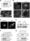
Src activation at the heterotypic contact during TEM of melanoma cells. (A) Immunoblots of stable transfectants expressing GFP-tagged WT and DN Src in WM239 melanoma cells. The endogenous Src is indicated by an arrow and GFP-tagged Src by an arrowhead. (B) Confocal image showing the localization of WT-Src-GFP in melanoma cell transfectants. (C) Confocal images showing the concentration of WT-Src-GFP at the heterotypic contact during both cell attachment and transmigration. Arrows indicate the heterotypic contact between the transfected melanoma cell and endothelial cells. (D) Immunolocalization of activated Src (p-Src) at the heterotypic contact during TEM of nontransfected melanoma cell. (a and b) Cocultures fixed at 1 h showing melanoma cells at the attachment stage. (c and d) Cocultures fixed at 5 h showing the transmigration stage. Control cocultures (a and c) and cocultures pretreated for 5 min with 1 mM pervanadate before fixation (b and d) were immunostained for p-Src. Melanoma cells are marked by an asterisk. Arrows indicate the heterotypic contact between melanoma cells and endothelial cells and arrowheads indicate the homotypic endothelial cell junction. Bars, 10 μm (B, C, and D). (E) Immunoblots showing that PP2 abolished Src activation during TEM. Melanoma cells were cultured on an endothelial monolayer in the presence or absence of PP2. Cells were collected at 0 or 5 h, and immunoblots were probed against p-Src and Src. (F) Immunoblots showing the p-Src level in WT-Src and DN-Src transfectants. (G) Src transfectants were cultured on an endothelial monolayer, and cells were collected at 0 or 5 h for immunoblot analysis of p-Src. In F and G, arrowheads indicate WT- or DN-Src-GFP, whereas arrows indicate the endogenous Src.
Figure 3.
Figure 3.
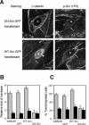
Effects of DN-Src expression on Src activation, β-catenin signaling, and TEM of melanoma cells. (A) Confocal images showing the immunolocalization of β-catenin and p-Src during TEM of Src-GFP transfectants (asterisks). Cells were treated with pervanadate for 5 min before immunostaining of p-Src. Arrows indicate the heterotypic contact between the Src-GFP transfectant and endothelial cells, and the arrowhead indicates the nuclear and perinuclear labeling of β-catenin. Bars, 10 μm. (B) Inhibition of β-catenin-mediated gene transcription in transmigrating melanoma cells by PP2 or DN-Src transfection. Cells were transiently transfected with a TOPflash vector and then cultured on top of an endothelial monolayer in the presence (▪) or absence (□) of PP2. Cells were collected at 5 h of coculture and assayed for luciferase activity. The luciferase activity at 5-h of coculture was normalized to that at 0-h coculture and the relative fold-increase in luciferase activity was calculated. Data represent the mean ± SD (n = 3). (C) Inhibition of TEM of melanoma cells by PP2 or DN-Src transfection. Cells were subjected to the TEM assay in the presence (▪) or absence (□) of PP2. The percentage of transmigrated cells was scored at 5 h. The data represent the mean ± SD (n = 3).
Figure 4.
Figure 4.
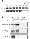
In vitro phosphorylation of the N-cadherin cytoplasmic domain disrupts its interaction with β-catenin. (A) Phosphorylation of recombinant N-cadherin cytoplasmic domain (N-cad-cyt) by an active Src. The phosphorylation reaction was carried out for different times, and aliquots of the reaction were subjected to SDS-PAGE. The immunoblots were probed with antibodies against N-cadherin and phospho-tyrosine. (B) Reduction of β-catenin binding to phosphorylated N-cadherin cytoplasmic domain (N-cad-cyt). GST-tagged N-cad-cyt was phosphorylated in vitro by Src in the presence or absence of ATP and then incubated with His-tagged β-catenin. The protein complex was isolated by glutathione beads, and immunoblots were probed for β-catenin, N-cadherin, and phospho-tyrosine.
Figure 5.
Figure 5.
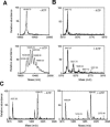
Identification of phospho-tyrosine residues on the N-cadherin cytoplasmic domain after in vitro phosphorylation by Src. The spectra are shown in pairs showing peptides that had been subjected to the phosphorylation reaction either in the presence or the absence of ATP. Phosphorylated peptides occurred as new peaks with an extra 80 Da in mass. (A) The His-tagged N-cadherin cytoplasmic domain was phosphorylated in vitro by Src and then subjected to MALDI-TOF mass spectrometry analysis. (B and C) His-tagged N-cadherin cytoplasmic domain was phosphorylated in vitro by Src and then digested by trypsin. The tryptic peptides were subjected to MALDI mass spectrometry analysis. (B) Identification of the phosphorylated Y852, Y860, Y884, and Y886 residues in the peptide AADNDPTAPPYDSLLVFDYEGSGSTAGSLSSLNSSSSGGEQDYDYLNDWGPR (5448 Da). (C) Identification of phospho-Y820 in peptides MDERPIHAEPQYPVR (1838 Da) and RMDERPIHAEPQYPVR (1994 Da).
Figure 6.
Figure 6.
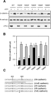
Effects of N-cadherin Y860 phosphorylation on β-catenin binding in vitro. (A) The five tyrosine residues identified on the N-cadherin cytoplasmic domain were mutated to phenylalanine individually. GST-tagged mutant proteins were purified from bacteria and then phosphorylated in vitro by Src. The phosphorylated proteins were subjected to the binding assay with His-tagged β-catenin. The protein complex was isolated by glutathione-Sepharose beads and the immunoblots were probed with antibodies against β-catenin, N-cadherin, and phospho-tyrosine. The phosphorylation of the protein carrying the Y860F mutation did not affect its binding with β-catenin (asterisk). (B) Quantification of the binding assays between mutant N-cadherin cytoplasmic domain and β-catenin. Before the binding assay, the mutant N-cadherin cytoplasmic domain was subjected to an in vitro Src phosphorylation reaction in the presence (black bars) or absence (gray bars) of ATP. The y-axis shows the ratio of bound β-catenin to N-cadherin cytoplasmic domain in the pull-down complex. *, Student's t test with p > 0.1, indicating that the level of β-catenin binding was not significantly different between the phosphorylated and the nonphosphorylated proteins. Data represent the mean ± SD (n = 3). (C) Conservation of the core β-catenin binding region in different classic cadherins. Tyr-852 and Tyr-860 of human N-cadherin and corresponding residues in the other cadherin sequences are shown in bold. The solid line indicates the core interacting region in the murine E-cadherin–β-catenin complex as identified by x-ray crystallography (Huber and Weis, 2001). The dash line indicates the core region identified in N-cadherin by peptide competition (Xu et al., 2002). *, the tyrosine residue essential for VE-cadherin–β-catenin interaction (Potter et al., 2005).
Figure 7.
Figure 7.
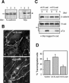
Effects of the expression of mutant N-cadherin on β-catenin dissociation and TEM of melanoma cells. (A) WM239 cells were transiently transfected with a myc-tagged wild-type or mutant (Y860F) N-cadherin. The expression of myc-tagged protein was confirmed by immunoblot analysis. The arrow indicates the endogenous N-cadherin, and the arrowhead indicates the expression of myc-tagged wild-type N-cadherin (lane 2) or myc-tagged mutant N-cadherin (lane 3). Control cells are shown in lane 1. (B) The transfectants were cocultured with an endothelial monolayer for 5 h before fixation and double staining using a myc mAb and a rabbit antibody against β-catenin. Myc staining was used to identify transfected cells (asterisks), and the confocal images show only β-catenin staining: melanoma cell expressing myc-tagged mutant N-cadherin (a) and melanoma cell expressing myc-tagged wild-type N-cadherin (b). Arrows indicate the heterotypic contact between transfectants and endothelial cells. Bars, 10 μm. (C) Melanoma cell were transiently transfected with wild-type N-cadherin-myc or mutant N-cadherin-myc and cocultured with an endothelial monolayer in a 3:1 ratio. Samples were collected at 0 or 5 h for immunoprecipitation with an antibody against myc. Protein blots of the immunoprecipitates were probed with antibodies against β-catenin, N-cadherin, and phospho-tyrosine. The pTyr-positive bands correspond to myc-tagged N-cadherin. (D) Transfected cells transiently expressing either wild-type or mutant myc-tagged N-cadherin were subjected to the TEM assay. Transmigration of myc-positive cells was scored at 5 h of coculture. Data represent the mean ± SD (n = 3).

References

    1. Behrens, J., Vakaet, L., Friis, R., Winterhager, E., Van Roy, F., Mareel, M. M., and Birchmeier, W. (1993). Loss of epithelial differentiation and gain of invasiveness correlates with tyrosine phosphorylation of the E-cadherin/β-catenin complex in cells transformed with a temperature-sensitive v-Src gene. J. Cell Biol. 120, 757–766. - PMC - PubMed
    1. Boyer, B., Bourgeois, Y., and Poupon, M. F. (2002). Src kinase contributes to the metastatic spread of carcinoma cells. Oncogene 21, 2347–2356. - PubMed
    1. Brabletz, T., Jung, A., Dag, S., Hlubek, F., and Kirchner, T. (1999). β-catenin regulates the expression of the matrix metalloproteinase-7 in human colorectal cancer. Am. J. Pathol. 155, 1033–1038. - PMC - PubMed
    1. Brabletz, T., Jung, A., Reu, S., Porzner, M., Hlubek, F., Kunz-Schughart, L. A., Knuechel, R., and Kirchner, T. (2001). Variable β-catenin expression in colorectal cancers indicates tumor progression driven by the tumor environment. Proc. Natl. Acad. Sci. USA 98, 10356–10361. - PMC - PubMed
    1. Brady-Kalnay, S. M., Rimm, D. L., and Tonks, N. K. (1995). Receptor protein tyrosine phosphatase PTPm associated with cadherins and catenins in vivo. J. Cell Biol. 130, 977–986. - PMC - PubMed

Publication types

MeSH terms