. 2006 Mar;17(3):1154-63.
doi: 10.1091/mbc.e05-04-0356. Epub 2005 Dec 21.

Barrier-to-autointegration factor phosphorylation on Ser-4 regulates emerin binding to lamin A in vitro and emerin localization in vivo

Affiliations

Barrier-to-autointegration factor phosphorylation on Ser-4 regulates emerin binding to lamin A in vitro and emerin localization in vivo

Luiza Bengtsson et al. Mol Biol Cell. 2006 Mar.

Abstract

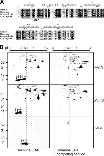
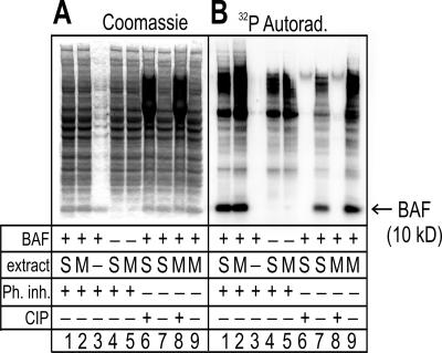
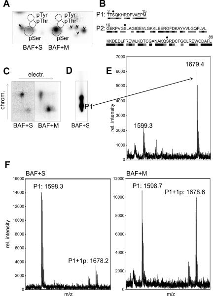
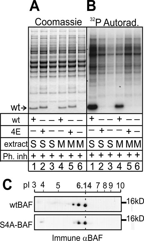
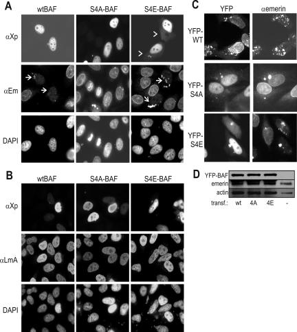
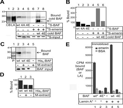
Barrier-to-autointegration factor (BAF) is a conserved 10-kDa chromatin protein essential in proliferating cells. BAF dimers bind double-stranded DNA, histone H3, histone H1.1, lamin A, and transcription regulators, plus emerin and other LEM-domain nuclear proteins. Two-dimensional gel analysis showed that endogenous human and Xenopus BAF are posttranslationally modified by phosphorylation and potentially other modifications and that they are hyperphosphorylated during mitosis. The invariant Ser-4 residue on BAF is a major site of phosphorylation during both interphase and mitosis. In HeLa cells that overexpressed the phosphomimetic BAF missense mutant S4E, but not S4A, emerin mislocalized from the nuclear envelope, suggesting Ser-4-nonphosphorylated BAF normally promotes emerin localization at the nuclear envelope. Supporting this model, wild-type BAF but not mutant S4E enhanced emerin binding to lamin A in vitro. Thus, Ser-4-unphosphorylated BAF has a positive role in localizing emerin; this role may be disease relevant because loss or mislocalization of emerin causes Emery-Dreifuss muscular dystrophy. Our findings further suggest Ser-4 phosphorylation inhibits BAF binding to emerin and lamin A, and thereby weakens emerin-lamin interactions during both mitosis and interphase.


Figures

Figure 1.
Figure 1.
Two-dimensional gel analysis of endogenous BAF. (A) Amino acid sequences of BAF from different species. The five α-helices (α1–5) and α-turn (“TT”) in human BAF are indicated. Serum 3273 antibodies were raised against residues 4–20 of human BAF (underlined). (B) Two-dimensionally (isoelectric focusing and SDS-PAGE) resolved HeLa cell and Xenopus egg extract proteins were immunoblotted with immune BAF serum 3273 (left) or immune serum plus competing peptide (right). M, M phase; S, S phase. Arrows and chevron indicate spots specifically recognized by serum 3273; chevron indicates unmodified BAF. Tilted and horizontal arrows indicate spots enriched in mitosis. The linear pI gradient is indicated above each set of panels. The vertical dotted line indicates pI 5.8, the calculated pI for unmodified human BAF.
Figure 2.
Figure 2.
Recombinant BAF is phosphorylated in both S- and M-phase Xenopus extracts. BAF dimers were incubated in S- or M-phase Xenopus extracts, and each entire reaction was resolved by SDS-PAGE and then Coomassie stained (A) and autoradiographed to detect incorporated 32P (B). M, M phase; Ph inh, phosphatase inhibitors; S, S phase.
Figure 3.
Figure 3.
Phosphoamino acid and mass spectrometric analysis of 10-kDa forms of BAF. (A) Phosphoamino acid analysis of phosphorylated BAF on TLC plates. Recombinant 10-kDa BAF incubated in Xenopus S- or M-phase extract was transferred to PVDF membrane, recovered from bands as in Figure 2, hydrolyzed, and resolved on TLC plates. Incorporated 32P was visualized by autoradiography, and phosphoamino acid standards (dotted circles) were visualized by ninhydrin staining. Chevrons indicate incomplete BAF hydrolysis products. (B) CNBr cleavage of BAF yields two peptides, P1 and P2. Serines are dotted. The predicted solvent accessibility of each residue, based on the crystal structure of the BAF dimer (Umland et al., 2000) is indicated by black (accessible), gray (intermediate), or white (buried). (C) The 32P-signal after thin layer electrophoresis and chromatography of CNBr-cleaved BAF. (D) The same fragments as in C but separated by chromatography only to maximize subsequent peptide recovery. (E) MALDI-MS spectrum of the peptide eluted from the P1 spot in D. (F) MALDI-MS spectra for P1 (mono- and unphosphorylated) in samples incubated in S-phase (BAF+S) or M-phase (BAF+M) Xenopus extracts.
Figure 4.
Figure 4.
Mutations at Ser-4 block phosphorylation of BAF in vitro and in vivo. Purified His-tagged BAF (wild type or S4E) were each incubated in S- or M-phase Xenopus egg extracts in the presence of [32P]ATP. Each whole reaction was then resolved by SDS-PAGE, Coomassie stained (A), and autoradiographed to detect incorporated 32P (B). M, M-phase; Ph inh, phosphatase inhibitors; S, S-phase. (C) Western blots of human kidney (293T) cells that were transiently transfected to overexpress either wild-type (WT) or S4A-mutant His/Xpress-tagged BAF and then separated on 2D gels, blotted and probed with BAF serum 3273. Dotted line indicates pI of unmodified His/Xpress-tagged BAF, which migrated at ∼16 kDa; endogenous BAF (10 kDa) is not shown.
Figure 5.
Figure 5.
Localization of endogenous emerin and A-type lamins in cells that overexpress wild-type or mutant BAF. (A and B) HeLa cells transiently transfected with cDNAs encoding either wild-type or mutant BAF were fixed and double stained by indirect immunofluorescence 19 h after transfection, using monoclonal antibodies against the N-terminal Xpress-tag (αXp) to visualize transfected BAF, and rabbit antibodies against emerin (A, αEm) or A-type lamins (B, αLmA). DNA was stained with 4,6-diamidino-2-phenylindole (DAPI). Chevrons in (A, top row) indicate cytoplasmic localization of BAF S4E protein. Arrows in (A, middle row) indicate emerin aggregates in cytoplasm. (C) HeLa cells expressing YFP-fused BAF (wild type, S4A, or S4E) stained by indirect immunofluorescence using antibodies against endogenous emerin (αEm). YFP-BAF was localized by YFP-autofluorescence. (D) Cells from C and control untransfected cells (–) were lysed and immunoblotted using antibodies against BAF, emerin, or actin.
Figure 6.
Figure 6.
Effect of BAF proteins (wild type and mutant) on the emerin–lamin A interaction. (A) Recombinant prelamin A tails immobilized on nitrocellulose membrane were probed with 35S-BAF, 35S-emerin, or 35S-emerin plus recombinant radioactive (cold) BAF. CB, strip stained with Coomassie blue. LA, strip probed with antibodies against lamin A. Bound nonradioactive recombinant BAF was detected using BAF serum 3273 (inset, lanes 7 and 8). Lowest panel (35S-input) shows 1% of each input 35S-labeled protein. (B) Densitometric quantification of results in A showing percentage of each probe bound, relative to the binding of 35S-emerin. (C) Nitro-cellulose-immobilized prelamin A tails were probed with His6-tagged bacterially expressed BAF that was preincubated with or without M-phase Xenopus extracts and then repurified on Ni2+-beads. Bound BAF was detected using BAF serum 3273. P, Ponceau S-stained lane to show prelamin A tail protein. (D) Densitometric quantification of results in C. Values are normalized to correct for different BAF input amounts. Binding of unmodified BAF was set as 100%. Results shown are typical of three repeats. (E) Recombinant emerin or BSA (control) were immobilized in microtiter wells and probed with 35S-BAF, 35S-prelamin A (full-length), or 35S-prelamin A (full length) plus recombinant radioactive (cold) BAF. Asterisks indicate 35S-labeled proteins.

Similar articles

Cited by

References

    1. Bengtsson, L., and Wilson, K. L. (2004). Multiple and surprising new functions for emerin, a nuclear membrane protein. Curr. Opin. Cell Biol. 16, 73–79. - PubMed
    1. Bradley, C. M., Ronning, D. R., Ghirlando, R., Craigie, R., and Dyda, F. (2005). Structural basis for DNA bridging by barrier-to-autointegration factor. Nat. Struct. Mol. Biol. 12, 935–936. - PubMed
    1. Broers, J. L., Hutchison, C. J., and Ramaekers, F. C. (2004). Laminopathies. J. Pathol. 204, 478–488. - PubMed
    1. Cai, M., Huang, Y., Zheng, R., Wei, S. Q., Ghirlando, R., Lee, M. S., Craigie, R., Gronenborn, A. M., and Clore, G. M. (1998). Solution structure of the cellular factor BAF responsible for protecting retroviral DNA from autointegration. Nat. Struct. Biol. 5, 903–909. - PubMed
    1. Casaday, R. J., Bailey, J. R., Kalb, S. R., Brignole, E. J., Loveland, A. N., Cotter, R. J., and Gibson, W. (2004). Assembly protein precursor (pU.L80.5 homolog) of simian cytomegalovirus is phosphorylated at a glycogen synthase kinase 3 site and its downstream “priming” site: phosphorylation affects interactions of protein with itself and with major capsid protein. J. Virol. 78, 13501–13511. - PMC - PubMed

Publication types

MeSH terms

LinkOut - more resources