Clinical Trial
. 2005 Dec 22;6(1):151.
doi: 10.1186/1465-9921-6-151.

MMP-9, TIMP-1 and inflammatory cells in sputum from COPD patients during exacerbation

Affiliations
Clinical Trial

MMP-9, TIMP-1 and inflammatory cells in sputum from COPD patients during exacerbation

P F Mercer et al. Respir Res. .

Abstract

Background: Irreversible airflow obstruction in Chronic Obstructive Pulmonary Disease (COPD) is thought to result from airway remodelling associated with aberrant inflammation. Patients who experience frequent episodes of acute deterioration in symptoms and lung function, termed exacerbations, experience a faster decline in their lung function, and thus over time greater disease severity However the mechanisms by which these episodes may contribute to decreased lung function are poorly understood. This study has prospectively examined changes in sputum levels of inflammatory cells, MMP-9 and TIMP-1 during exacerbations comparing with paired samples taken prior to exacerbation.

Methods: Nineteen COPD patients ((median, [IQR]) age 69 [63 to 74], forced expiratory volume in one second (FEV1) 1.0 [0.9 to 1.2], FEV1% predicted 37.6 [27.3 to 46.2]) provided sputa at exacerbation. Of these, 12 were paired with a samples collected when the patient was stable, a median 4 months [2 to 8 months] beforehand.

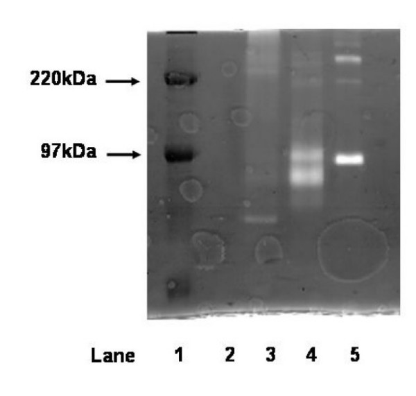
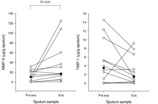
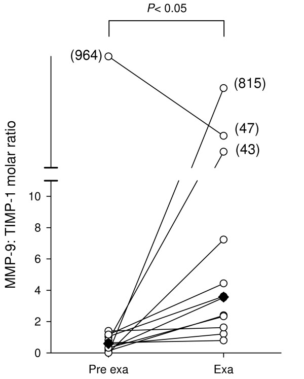
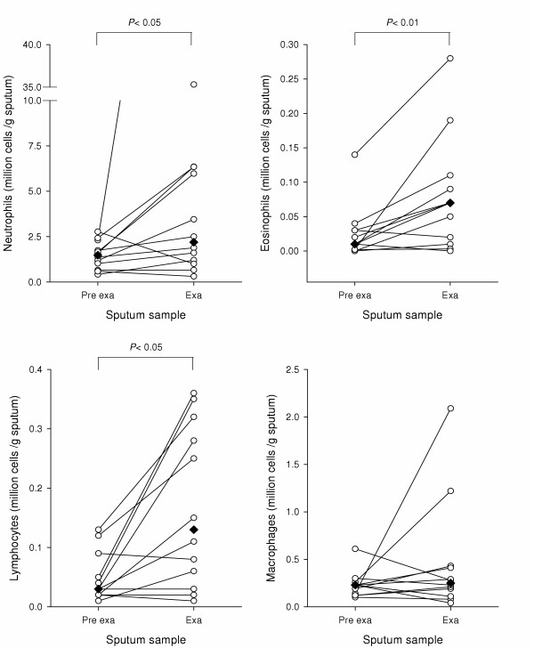
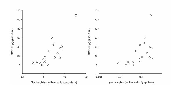
Results: MMP-9 levels increased from 10.5 microg/g [1.2 to 21.1] prior to exacerbation to 17.1 microg/g [9.3 to 48.7] during exacerbation (P < 0.01). TIMP-1 levels decreased from 3.5 microg/g [0.6 to 7.8] to 1.5 microg/g [0.3 to 4.9] (P = 0.16). MMP-9/TIMP-1 Molar ratio significantly increased from 0.6 [0.2 to 1.1] to 3.6 [2.0 to 25.3] (P < 0.05). Neutrophil, eosinophil and lymphocyte counts all showed significant increase during exacerbation compared to before (P < 0.05). Macrophage numbers remained level. MMP-9 levels during exacerbation showed highly significant correlation with both neutrophil and lymphocyte counts (Rho = 0.7, P < 0.01).

Conclusion: During exacerbation, increased inflammatory burden coincides with an imbalance of the proteinase MMP-9 and its cognate inhibitor TIMP-1. This may suggest a pathway connecting frequent exacerbations with lung function decline.


Figures

Figure 1
Figure 1
Sample zymogram showing immunoprecipitation of MMP-9 from sputum. 10 μl volumes loaded per lane. Lane 1: High range molecular weight markers (Amersham Biosciences); lane 2: polyclonal antiMMP-9 (4 μg/ml); lane 3: exacerbation sputum sample (1:100 dilution) + antiMMP-9 (4 μg/ml); lane 4: exacerbation sputum sample (1:100 dilution); lane 5: pro-MMP-9 standard (92 kDa) (100 ng/ml).
Figure 2
Figure 2
MMP-9 and TIMP-1 levels in sputum from COPD patients. Samples were taken from patients prior to and during exacerbation (n = 12). MMP-9 was measured by gelatin zymography and TIMP-1 was measured by ELISA and corrected for weight of sputum. Individuals are shown as open circles and median values as diamonds. Data were analysed by non parametric Wilcoxon signed rank test.
Figure 3
Figure 3
Molar ratio between MMP-9 and TIMP-1 in COPD sputum. The data from figure 2 was used to calculate a molar ratio between MMP-9 and TIMP-1. Median values are marked as diamonds. Data were statistically analysed by a non parametric Wilcoxon signed rank test.
Figure 4
Figure 4
Inflammatory cell counts prior to and during exacerbation. Differential neutrophil, eosinophil, lymphocyte and macrophage counts were carried out on samples from COPD patients and corrected for weight of sputum. Samples were taken from patients prior to and during exacerbation (n = 12). Median cell counts are marked as diamonds. Data were analysed by a non parametric Wilcoxon signed rank test.
Figure 5
Figure 5
Relationship between cell infiltration and MMP-9 in COPD sputum during an exacerbation. Neutrophil (Left hand panel) and lymphocyte (right hand panel) counts were carried out on sputum samples from COPD patients during exacerbation (n = 19), and corrected for weight of sputum. Measurement of MMP-9 levels was carried out on cell free sputum supernatants. Cell count data were expressed as million cells/g (log scale) and MMP-9 data as μg/g.

Similar articles

Cited by

References

    1. Hogg JC, Senior RM. Chronic obstructive pulmonary disease - part 2: pathology and biochemistry of emphysema. Thorax. 2002;57:830–834. doi: 10.1136/thorax.57.9.830. - DOI - PMC - PubMed
    1. Atkinson JJ, Senior RM. Matrix metalloproteinase-9 in lung remodeling. Am J Respir Cell Mol Biol. 2003;28:12–24. doi: 10.1165/rcmb.2002-0166TR. - DOI - PubMed
    1. Barnes PJ. New treatments for COPD. Nat Rev Drug Discov. 2002;1:437–446. doi: 10.1038/nrd820. - DOI - PubMed
    1. Visse R, Nagase H. Matrix metalloproteinases and tissue inhibitors of metalloproteinases: structure, function, and biochemistry. Circ Res. 2003;92:827–839. doi: 10.1161/01.RES.0000070112.80711.3D. - DOI - PubMed
    1. Betsuyaku T, Nishimura M, Takeyabu K, Tanino M, Venge P, Xu S, Kawakami Y. Neutrophil granule proteins in bronchoalveolar lavage fluid from subjects with subclinical emphysema. Am J Respir Crit Care Med. 1999;159:1985–1991. - PubMed

Publication types

Substances