. 2006 Mar 1;571(Pt 2):303-17.
doi: 10.1113/jphysiol.2005.100719. Epub 2005 Dec 22.

ATP-sensitive K+ channels: regulation of bursting by the sulphonylurea receptor, PIP2 and regions of Kir6.2

Affiliations

ATP-sensitive K+ channels: regulation of bursting by the sulphonylurea receptor, PIP2 and regions of Kir6.2

Bernard Ribalet et al. J Physiol. .

Abstract

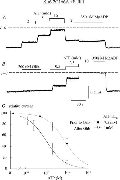
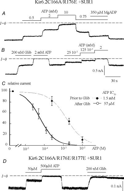
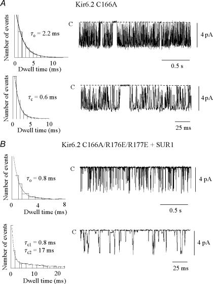
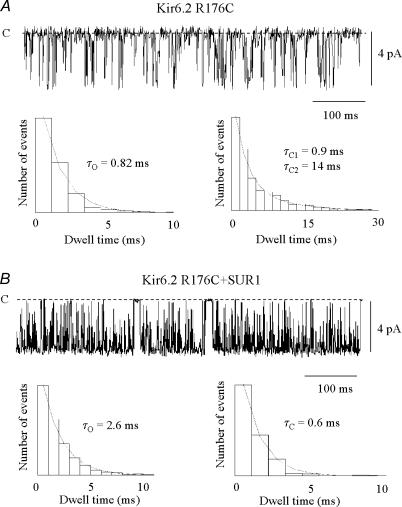
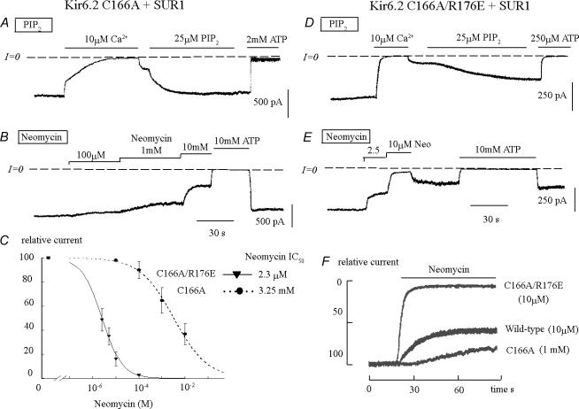
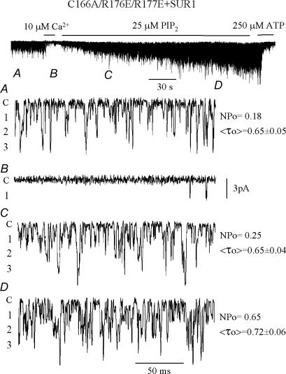
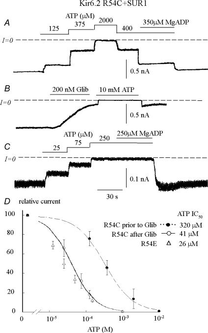
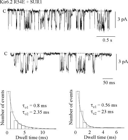
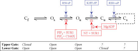
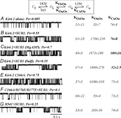
ATP-sensitive K+ channels composed of the pore-forming protein Kir6.2 and the sulphonylurea receptor SUR1 are inhibited by ATP and activated by Phosphatidylinositol Bisphosphate (PIP2). Residues involved in binding of these ligands to the Kir6.2 cytoplasmic domain have been identified, and it has been hypothesized that gating mechanisms involve conformational changes in the regions of the bundle crossing and/or the selectivity filter of Kir6.2. Regulation of Kir6.2 by SUR1, however, is not well-understood, even though this process is ATP and PIP2 dependent. In this study, we investigated the relationship between channel regulation by SUR1 and PIP2 by comparing a number of single and double mutants known to affect open probability (P(o)), PIP2 affinity, and sulphonylurea and MgADP sensitivity. When coexpressed with SUR1, the Kir6.2 mutant C166A, which is characterized by a P(o) value close to 0.8, exhibits no sulphonylurea or MgADP sensitivity. However, when P(o) was reduced by combining mutations at the PIP2-sensitive residues R176 and R177 with C166A, sulphonylurea and MgADP sensitivities were restored. These effects correlated with a dramatic decrease in PIP2 affinity, as assessed by PIP2-induced channel reactivation and inhibition by neomycin, an antagonist of PIP2 binding. Based on macroscopic and single-channel data, we propose a model in which entry into the high-P(o) bursting state by the C166A mutation or by SUR1 depends on the interaction of PIP2 with R176 and R177 and, to a lesser extent, R54. In conjunction with this PIP2-dependent process, SUR1 also regulates channel activity via a PIP2-independent, but MgADP-dependent process.


Figures

Figure 1
Figure 1. ATP and MgADP sensitivity of C166A mutant channels coexpressed with SUR1 before and after addition of glibenclamide
Current traces in the two upper panels depict inward currents measured before (A) and after (B) application of 200 nm glibenclamide. Currents were progressively suppressed by increasing ATP concentrations, with the half-maximal inhibition (IC50) approximately 7.5 mm and 1 mm, respectively. The main observations are that C166A + SUR1 channels are poorly sensitive to MgADP (A) and the sulphonylurea glibenclamide (B). However, a small change in Po due to glibenclamide (B) causes a dramatic increase in ATP sensitivity. C, graph showing ATP sensitivity for C166A + SUR1 channels before (•) and after (○) application of glibenclamide. Fit of the data points to the Hill equation yielded IC50 values of 7.5 mm (n = 5) before and 1 mm (n = 4) after addition of glibenclamide. In both cases the Hill coefficients were close to unity.
Figure 2
Figure 2. Properties of C166A/R176E (AC) and C166A/R176E/R177E channels (D) coexpressed with SUR1 before and after addition of glibenclamide
Current traces in the two upper panels depict inward currents from C166A/R176E + SUR1 channels measured before (A) and after (B) application of 200 nm glibenclamide. Currents were progressively suppressed by increasing ATP concentrations, with IC50 values near 1.5 mm and 50 μm, respectively. In contrast to C166A + SUR1 channels (Fig. 1), C166A/R176E + SUR1 channels demonstrate high sensitivity to MgADP (A) and the sulphonylurea glibenclamide (B). Like C166A + SUR1 channels (Fig. 1), exposure to glibenclamide causes a dramatic increase in ATP sensitivity. C, graph showing ATP sensitivity for C166A/R176E + SUR1 channels before (•) and after (○) application of glibenclamide. Fit of the data points to the Hill equation yielded IC50 values of 1.5 mm (n = 4) before and 57 μm (n = 3) after application of glibenclamide. The two Hill coefficients were close to unity. D, inward current recording from C166A/R176E/R177E + SUR1 channels. The current amplitude was greatly diminished as compared to A and B, and inhibition by glibenclamide exceeded 90%. The channel sensitivity to ATP was similar to C166A-R176E + SUR1 channels after addition of glibenclamide, with an IC50 of 45 μm.
Figure 3
Figure 3. Single channel recordings from C166A and C166A/R176E/R177E channels
A, inward currents obtained with the C166A mutant were recorded in excised inside-out patches at a holding potential of −60 mV and are represented as downward deflections, showing bursts of openings. The lower trace shows single-channel recording on an expanded timescale. The open-time histogram on the top left was well-fitted with a single exponential (τo= 2.2 ms). The closed-time histogram below required two exponentials (τc1= 0.6 ms and τc2= 15 ms). However, the slow (interburst) time constant component contributed little to the total number of events (see Table 1). In contrast, C166A/R176E/R177E channels opened only with short openings and demonstrated a fast gating behaviour (B) similar to wild-type Kir6.2 channels in the absence of SUR1. This behaviour is illustrated in the expanded lower single-channel current tracing. In this case the open-time histogram on the left was fitted by a single exponential (τo= 0.8 ms), but a second slower time constant could also be observed (Table 1). The closed-time histogram below also required two exponentials (τc1= 0.8 ms and τc2= 17 ms).
Figure 4
Figure 4. Single-channel recordings from R176C channels without and with SUR1
A, inward currents obtained with the R176C mutant were recorded in the absence of SUR1 in excised inside-out patches at a holding potential of −60 mV and are represented as downward deflections, showing brief openings. In this case the channel Po was 0.13. The open-time histogram in the bottom left panel was fitted with a single exponential (τo= 0.82 ms), while the closed-time histogram on the right required two exponentials (τc1= 0.9 ms and τc2= 14 ms). In contrast, in the presence of SUR1, R176C channels opened in a long burst (B). In this case channel Po approached maximum (0.8). The open-time histogram in the bottom left panel was fitted by a single exponential (τo= 2.6 ms) and the closed-time histogram on the right also required one exponential (τc= 0.6 ms).
Figure 5
Figure 5. PIP2 and neomycin sensitivity of Kir6.2 C166A + SUR1 and Kir6.2 C166A/R176E + SUR1 channels coexpressed with SUR1
Current traces in A and D illustrate the reactivation of Kir6.2 C166A and Kir6.2 C166A/R176E channels by PIP2, respectively. A, the activity of C166A + SUR1 was first blocked by addition of Ca2+. Upon removal of Ca2+, there was partial recovery of channel activity; the level of recovery was dependent upon the duration of Ca2+ exposure. Addition of PIP2 under these conditions caused full recovery of channel activity within 30 s. D, the activity of C166A/R176E + SUR1 was first blocked with Ca2+; upon addition of PIP2 there was only partial and slow recovery of channel activity consistent with low PIP2 affinity. Also consistent with low PIP2 affinity of C166A/R176E, we found that hydrolysis of PIP2, as illustrated by Ca2+-dependent rate of channel block, was much faster with the double mutant C166A/R176E + SUR1 as compared to with C166A + SUR1. data in B and E show how neomycin may be used to assess this difference in PIP2 affinity and graphs in C and F illustrate how the effect of neomycin may be quantified. These data illustrate the potent increase in neomycin sensitivity elicited by R176E, which parallels the weak channel reactivation by PIP2 in D. Consistent with the low PIP2 affinity of C166A/R176E, 10 mm MgATP could restore only partially the activity of this channel (E), while reactivating fully C166A (B). C, fitting of the neomycin data points to the Hill equation yielded IC50 values of 3.25 mm (n = 4) for C166A + SUR1 (•) and 2.3 μm (n = 4) for C166A/R176E + SUR1 (♦). In both cases the Hill coefficients were less than unity. F, the rate of inhibition by neomycin is compared for C166A + SUR1 and C166A/R176E + SUR1 channels. In this example 10 μm neomycin caused 50% inhibition of C166A/R176E + SUR1 within 4 s, while 1 mm neomycin caused only gradual and partial inhibition of C166A + SUR1 over several minutes. The rate of inhibition of wild-type Kir6.2 channel by 10 μm neomycin is shown for comparison.
Figure 6
Figure 6. Effects on single channel kinetics of reactivation of C166A/R176E/R177E channels by PIP2 in the absence of Mg2+
Upper trace, inward currents obtained with the C166A/R176E/R177E mutant were recorded in excised inside-out patches at a holding potential of −60 mV and are represented as downward deflections. This recording illustrates the gradual increase in channel activity elicited by PIP2 and the block by ATP. The lower four traces show single channel recording on an expanded timescale, before (A) and after application of PIP2 (C and D). The values on the right of the traces illustrate the lack of effect of PIP2 on the mean channel open time (compare A and C), thus, the increase in channel activity is probably due to channel recruitment. The mean open time was measured in all cases at the first level of opening and due to the presence of multiple channels in the patch the value of <τo > is not an accurate measurement of single-channel open time, but represents a compound value. The modest increment in <τo > in D reflects increased simultaneous opening of two or more channels.
Figure 7
Figure 7. ATP and MgADP sensitivity of R54C and R54E channels coexpressed with SUR1, before and after addition of glibenclamide
Current traces depict inward currents measured before (A) and after (B and C) application of 200 nm glibenclamide. Currents were progressively suppressed by increasing ATP concentrations, with the IC50 values near 320 μm and 40 μm, respectively. R54C + SUR1 channels were reactivated by MgADP (A) and inhibited by the sulphonylurea glibenclamide (B). D, graph showing ATP sensitivity for R54C + SUR1 channels before (•) and after (○) application of glibenclamide. Fit of the data points to the Hill equation yielded IC50 values of 320 μm (n = 5) before and 41 μm (n = 5) after application of glibenclamide. In both cases the Hill coefficients were close to unity. D shows R54C + SUR1 data as well as data obtained with R54E + SUR1 channels. The IC50 for ATP of R54E + SUR1 channels was recorded after channel run-down. The small increase in ATP sensitivity from 41 to 26 μm with R54C and R54E, respectively, is likely to be due to the decreased Po of R54E + SUR1 compared to R54C + SUR1 channels.
Figure 8
Figure 8. Single-channel recordings from R54E + SUR1 channels
Inward currents were recorded in excised inside-out patches at a holding potential of −60 mV and are represented as downward deflections. The lower record shows data on an expanded timescale. The open-time histogram on the left was fitted with a single exponential (τo= 2.3 ms). The closed-time histogram required two exponentials (τc1= 0.6 ms and τc2= 23 ms). The relative contribution of these two components is given in Table 1.
Figure 9
Figure 9. Schematic diagram of channel regulation by ATP, SUR, MgADP and PIP2 based on a five-state model
The model is based on a previously proposed minimal model to account for adenine nucleotide sensitivity of Kir6.2 + SUR1 (John et al. 2003), with putative interactions of the α, β and γ phosphate groups of ATP with Kir6.2 residues indicated in blue. The ATP-bound states and the Cs state used previously are not included in the present modelling because the experiments were carried out in the absence of ATP; in the absence of ATP the Cs state is almost never visited. We incorporate into this model the regulatory effect of SUR1, PIP2, MgADP and sulphonylureas as follows. In the absence of SUR1 or C166A, the channel transitions with fast kinetics between the Ou and Cu states, in which the lower gate (near the bundle-crossing or G-loops) is unstable. In the presence of SUR1 or when C166 is mutated to C166A/S, channel interaction with PIP2 stabilizes the lower gate, so that the channel enters the bursting states Os and Cf, in which opening and closing transitions are mediated by the upper gate (at the selectivity filter). A second effect of SUR1, which involves the inhibitory effect of the N-terminus on channel gating, takes place at the Cu to Ou transition, such is relieved when MgADP binds to SUR1, thus, causing maximal channel activation. The effect of MgADP is inhibited by sulphonylureas, decreasing channel Po. Simulations related to this model are shown in Fig. 10.
Figure 10
Figure 10. Simulation of single channel kinetic behaviour using the model in Fig. 9
The numbers in the reaction scheme at the top indicate rate constants (in units of s−1) that were fixed throughout the simulations. The values for the Os to Cf and Cf to Os transitions were obtained using C166A mutant data and are similar to those published by other investigators (see Appendix). The values for the other rate constants (KOsOu, KOuOs and KCuOu), which were fitted to simulate different conditions in AG, are shown in italics at the right of each simulated single-channel current tracing. Fitting to the model shown at the top was carried out using the MIL function of the QuB software (University at Buffalo, NY, USA). Interventions in A and EG could be fitted by changes of one rate constant, KOuOs, reflecting the channel's affinity for PIP2. In BD, illustrating the effects of SUR1 (B), SUR1 + MgADP (C) and SUR1 + sulphonylurea (D), all the data could be fitted by changes of the KCuOu value (bold characters). Channel Po values were calculated for each experimental condition and are indicated above each single channel current trace. See Discussion and Appendix for further details.

Similar articles

Cited by

References

    1. Alekseev AE, Brady PA, Terzic A. Ligand-insensitive state of cardiac ATP-sensitive K+ channels. Basis for channel opening. J Gen Physiol. 1998;111:381–394. - PMC - PubMed
    1. Babenko AP, Bryan J. Sur domains that associate with and gate KATP pores define a novel gatekeeper. J Biol Chem. 2003;278:41577–41580. - PubMed
    1. Babenko AP, Gonzalez G, Bryan J. The N-terminus of KIR6.2 limits spontaneous bursting and modulates the ATP-inhibition of KATP channels. Biochem Biophys Res Commun. 1999;255:231–238. - PubMed
    1. Baukrowitz T, Schulte U, Oliver D, Herlitze S, Krauter T, Tucker SJ, Ruppersberg JP, Fakler B. PIP2 and PIP as determinants for ATP inhibition of KATP channels. Science. 1998;282:1141–1144. - PubMed
    1. Drain P, Li L, Wang J. KATP channel inhibition by ATP requires distinct functional domains of the cytoplasmic C terminus of the pore-forming subunit. Proc Natl Acad Sci U S A. 1998;95:13953–13958. - PMC - PubMed

Publication types

MeSH terms

LinkOut - more resources