Review
. 2006 Jan;15(1):1-13.
doi: 10.1110/ps.051658406.

Breaking symmetry in protein dimers: designs and functions

Affiliations
Review

Breaking symmetry in protein dimers: designs and functions

Jerry H Brown. Protein Sci. 2006 Jan.

Abstract

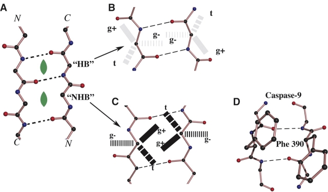
Symmetry, and in particular point group symmetry, is generally the rule for the global arrangement between subunits in homodimeric and other oligomeric proteins. The structures of fragments of tropomyosin and bovine fibrinogen are recently published examples, however, of asymmetric interactions between chemically identical chains. Their departures from strict twofold symmetry are based on simple and generalizable chemical designs, but were not anticipated prior to their structure determinations. The current review aims to improve our understanding of the structural principles and functional consequences of asymmetric interactions in proteins. Here, a survey of >100 diverse homodimers has focused on the structures immediately adjacent to the twofold axis. Five regular frameworks in alpha-helical coiled coils and antiparallel beta-sheets accommodate many of the twofold symmetric axes. On the basis of these frameworks, certain sequence motifs can break symmetry in geometrically defined manners. In antiparallel beta-sheets, these asymmetries include register slips between strands of repeating residues and the adoption of different side-chain rotamers to avoid steric clashes of bulky residues. In parallel coiled coils, an axial stagger between the alpha-helices is produced by clusters of core alanines. Such simple designs lead to a basic understanding of the functions of diverse proteins. These functions include regulation of muscle contraction by tropomyosin, blood clot formation by fibrin, half-of-site reactivity of caspase-9, and adaptive protein recognition in the matrix metalloproteinase MMP9. Moreover, asymmetry between chemically identical subunits, by producing multiple equally stable conformations, leads to unique dynamic and self-assembly properties.


Figures

Figure 1.
Figure 1.
Register asymmetry in a dimeric antiparallel β-sheet. (A) An antiparallel β-sheet (depicted schematically) composed of two chemically identical chains may be twofold symmetric when analogous positions along the amino acid sequence (e.g., i and i′) are located across from each other at the dimer axis; here, the midpoint (red circle) between analogous residues of the dimer coincides with a local twofold symmetry axis (hollow black oval) accorded by the geometry of the main chain (black). (Note that analogous residues in a symmetrical register may either be main-chain “hydrogen-bonded” to each other [as pictured here] or form a “non-hydrogen-bonded” pair as explained in Fig. 2; see also Table 1). (B) When the register of one of the chains is “shifted” by one position (or any odd number of positions) relative to that in A, only different positions along the amino acid sequence (e.g., i and i i′ – 1) are located directly across from each other; in this case the midpoint (red circle) between analogous residues (e.g., i and i′) of the dimer falls on a non-twofold symmetric location of the main-chain structure (i.e., the red circle does not coincide with a hollow black oval). Some of the chemical consequences of this register shift for any pair of analogous residues are that one of their main-chain carbonyls points into the dimer interface while the other points away, and that their side chains are found on opposite faces of the β-sheet (here, i [bold red] faces toward the reader and i′ [faded light red] faces away; see also E). (Note that in the language of Fig. 2, residue i in this asymmetric register is forming main-chain hydrogen bonds across the dimer interface [to i′ – 1], while the analogous residue i′ is not.) (C) In the N-terminal γ domain of the dimeric bovine fibrinogen molecule (Madrazo et al. 2001), the asymmetrically registered alignment is enforced in an antiparallel β-sheet-like structure by a rare arrangement of consecutive disulfides that covalently link residue γ8 (i) to residue γ′ 9 (i′ + 1), as well as γ′ 8 to γ9. The asymmetry of this arrangement is propagated throughout the N-terminal 14 residues of the two γ chains and may have important implications for the uniformity of the fibrin clot. (We also note that a pair of adjacent reciprocal disulfide bonds connects the subunits of seminal bovine ribonuclease [PDB 11BG], but in this case they are part of α-helices and the dimer is roughly symmetric.) (D,E) The dimeric interface of mannose-binding protein can adopt either the symmetric (D) or asymmetric (E) register, depending upon crystallization conditions (Weis et al. 1991, 1992). Note that the side-chain interactions between the strands nearest the central axis of the dimer are similar in the two cases, due to chemical similarity (common γ methyl group) of the consecutive valine and threonine side chains (see text). Other interactions (not shown) more distant from the central axis differ between the two crystal forms. (F) For antiparallel strands consisting only of alternating charged residues (e.g., the displayed polar zipper of Perutz and colleagues [De Baere et al. 1992; Perutz 1994]), a symmetrical register necessarily yields a residue pair of like charges at the twofold axis. (G) For such a peptide, a complementary network of oppositely charged residues can only be produced in an asymmetric register.
Figure 2.
Figure 2.
Side-chain rotamer asymmetry in a dimeric antiparallel β-sheet. (A) An antiparallel β-pleated sheet may accommodate a perpendicularly oriented twofold axis (green filled oval) in one of two geometrically distinctive locations: at the center of a pair of residues that are hydrogen bonded to each other (HB) or at the center of a pair of residues that are not hydrogen bonded to each other (NHB). (B,C) On each of the two residue-pair geometries, these figures superimpose the three staggered rotameric conformations (about the Cα–Cβ bond) that are most commonly observed for side chains. In proteins in general, the relative populations of these rotamers to some extent depend both on the identity of the side chain and the φ–ψ angles of the main chain (Dunbrack and Karplus 1993), but usually the g+ rotamer is most favored. (B) For an HB residue pair, two side chains both having the g+ rotameric conformation face away from one another. (C) For the NHB pair, such side chains are in position to interact most closely with one another. (D) In caspase-9 (Renatus et al. 2001), the dimer axis is located at the center of an NHB pair of phenylalanines (390 and 390′). These bulky side chains would clash if they both adopted the g+ conformation, specifically because of the NHB geometry at the dimer axis. The asymmetry that in this case occurs (where one phenylalanine adopts the g+ conformation and the other is swung away) is propagated throughout the respective monomers (see Renatus et al. 2001) and apparently causes the dimer to display half-site reactivity (Renatus et al. 2001). Similar asymmetry occurs in insulin dimers at Phe25 (not shown).
Figure 3.
Figure 3.
Two types of locations accommodate a twofold rotation axis in antiparallel α-helical coiled coils. The number of residues in each chain that span consecutive core positions alternate between four (the a, b, c, and d positions) and five (the d, e, f, g, and a positions); the two-stranded coiled coil may thus be visualized as consisting of alternating “b-centered” ((abcd)2) and “f-centered” ((defga)2) rings of residues. Similar to the alternating hydrogen-bonded and non-hydrogen-bonded residue pairs in the antiparallel β-sheets, the b-centered and f-centered rings are geometrically distinctive, and in antiparallel coiled coils each of them can accommodate a perpendicularly oriented dyad at its center (green filled ovals).
Figure 4.
Figure 4.
Axial-shift asymmetry in parallel α-helical coiled coils. (A) Parallel α-helical coiled coils may accommodate a twofold rotation axis running along the length of the coiled-coil axis (green double-headed arrow). (B) Symmetry is broken when the two chains are axially out of register. (C) In tropomyosin (and other proteins), a relatively symmetrical coiled-coil segment occurs with core leucines that fit into four-residue holes of the adjacent helix whereas (D) local axial staggering is promoted by core alanines (Brown et al. 2001) because of their preferential fit into axially shifted three-residue holes (Gernert et al. 1995; Walther et al. 1996).
Figure 5.
Figure 5.
Junction bends in asymmetric homo-oligomers. The junction of symmetric (bottom half) and asymmetric (upper half) segments of a protein can predictably control the molecule’s overall shape. In a parallel dimeric molecule (left) such as tropomyosin, cortexillin, or the DNA-binding protein PUT3 (see text; Brown et al. 2001), a relatively small (~1.2 Å) axial staggering of relatively widely separated (8–10 Å) α-helices yields a small wedge (triangle) in one chain away from which the molecular axis (solid lines) thus bends by a small (~3–6) degree. This geometrical design applies to the trimeric collagen–foldon structure (Stetefeld et al. 2003) (right), where the relatively large (~5–6 Å) axial stagger between narrowly separated (~4–5 Å) collagen chains leads to a severe (~63°) bend (see text).
Figure 6.
Figure 6.
Conformational degeneracy in asymmetric homodimers. (A) When an object dimerizes asymmetrically, the choice of one subunit, say the left one, adopting conformation “X” and the right subunit adopting conformation “X′” would be energetically indistinguishable from the alternative of the left subunit adopting conformation X′ and the right one adopting conformation X. The rate of interchange between these degenerate states likely varies from case to case (see text for description of tropomyosin [“Tm”] and fibrinogen [“Fgen”] structures). (B) Piston-like motions between two equivalently stabilized pairs of axially staggered chains in a parallel homodimer. (C) Bistable joints in a homodimer containing symmetrically in-register (red) and asymmetrically staggered (blue) segments (such as tropomyosin). (D) Irregular self-assemblies arising from asymmetric contacts (such as in the protofilament backbone of fibrin displayed here schematically; see “Nonuniformities in self-assembly” section of text for details).

References

    1. Amor, J.C., Harrison, D.H., Kahn, R.A., and Ringe, D. 1994. Structure of the human ADP-ribosylation factor 1 complexed with GDP. Nature 372: 704–708. - PubMed
    1. Anfinsen, C.B. 1973. Principles that govern the folding of protein chains. Science 181: 223–230. - PubMed
    1. Banner, D.W., Kokkinidis, M., and Tsernoglou, D. 1987. Structure of the ColE1 rop protein at 1.7 Å resolution. J. Mol. Biol. 196: 657–675. - PubMed
    1. Berger, B., Shor, P.W., Tucker-Kellogg, L., and King, J. 1994. Local rule-based theory of virus shell assembly. Proc. Natl. Acad. Sci. 91: 7732–7736. - PMC - PubMed
    1. Brown, J.H., Volkmann, N., Jun, G., Henschen-Edman, A.H., and Cohen, C. 2000. The crystal structure of modified bovine fibrinogen. Proc. Natl. Acad. Sci. 97: 85–90. - PMC - PubMed

Publication types