. 2006 Jan;12(1):94-101.
doi: 10.1261/rna.2264806.

Virus-like particles of the Ty3 retrotransposon assemble in association with P-body components

Affiliations

Virus-like particles of the Ty3 retrotransposon assemble in association with P-body components

Nadejda Beliakova-Bethell et al. RNA. 2006 Jan.

Abstract

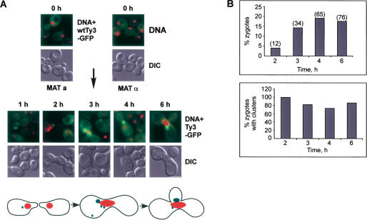
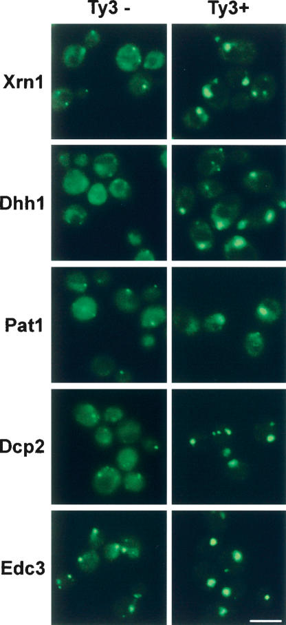
Retroviruses and retrotransposons assemble intracellular immature core particles around a RNA genome, and nascent particles collect in association with membranes or as intracellular clusters. How and where genomic RNA are identified for retrovirus and retrotransposon assembly, and how translation and assembly processes are coordinated is poorly understood. To understand this process, the subcellular localization of Ty3 RNA and capsid proteins and virus-like particles was investigated. We demonstrate that mRNAs, proteins, and virus-like particles of the yeast Ty3 retrotransposon accumulate in association with cytoplasmic P-bodies, which are sites of mRNA translation repression, storage, and degradation. Deletions of genes encoding P-body proteins decreased Ty3 transposition and caused changes in the pattern of Ty3 foci, underscoring the biological significance of the association of Ty3 virus-like protein components and P-bodies. These results suggest the hypothesis that P-bodies may serve to segregate translation and assembly functions of the Ty3 genomic RNA to promote assembly of virus-like particles. Because Ty3 has features of a simple retrovirus and P-body functions are conserved between yeast and metazoan organisms, these findings may provide insights into host factors that facilitate retrovirus assembly.


Figures

FIGURE 1.
FIGURE 1.
Ty3-GFP, similar to wild-type Ty3, produces clusters of VLPs. (A) Ty3-GFP/RFP in pNB2127/pNB2266. The GFP/RFP coding region was inserted at an XmaI site so that GFP/RFP was expressed fused to the POL3 reading frame at the downstream end. (B) Cells expressing Ty3-GFP/RFP show correct proteolytic maturation of Gag3 into p27 and CA and production of IN-GFP/RFP from Gag3-Pol3-GFP/RFP. Cultures of BY4741 transformed with pDLC201, pNB2127, or pNB2266 were induced for Ty3 or Ty3-GFP/RFP expression for 6 h. Cell extracts were prepared and fractionated by electrophoresis on SDS–10% polyacrylamide gels. Proteins were transferred to Immobilon and analyzed by reacting with anti-GFP, anti-IN, and anti-CA IgG. Uninduced cell extracts are indicated by “U.” Positions of IN-GFP/RFP (88 kDa), IN (61 kDa), Gag3 (34 kDa), and CA (24 kDa) are indicated. (C) BY4741 cells transformed with pNB2127 and induced to express Ty3-GFP for different periods of time. Ty3-GFP is indicated by green; DAPI-stained DNA, pseudo-colored red. Scale bar, 5.0 μm. (D) BY4741 cells transformed with pNB2127 and induced for 6 h to express Ty3-GFP observed by EM. Scale bar, 0.5 μm; n indicates nucleus; v, vacuole. (E) Cells induced for Ty3 expression as described in D, except processed by reacting sequentially with rabbit polyclonal anti-GFP and goat anti-rabbit IgG conjugated to 15-nm gold particles. Scale bar, 0.24 μm.
FIGURE 2.
FIGURE 2.
Ty3 proteins occur in foci when Ty3 is expressed under its native promoter in mating cells. Sequences containing the coding region of Ty3-GFP were cloned under the native pheromone-inducible Ty3 LTR in the low-copy vector pRS316 (pNB2176). Ty3 BY4741 (MATa) cells transformed with pNB2176 and BY4742 (MATα) cells transformed with the high-copy vector pYES2.0 were mixed on solid SD-ura and allowed to undergo mating to induce Ty3-GFP expression. (A) Ty3-GFP clusters form in mating cells. Formation of zygotes at 0, 1, 2, 3, 4, and 6 h after mixing. (Top, upper panels) Nuclei and Ty3 clusters (DAPI, red; Ty3-GFP, green). (Top, lower panels) Differential interference contrast (DIC) micrographs of whole cells. Scale bar, 5.0 μm. (Bottom) Cartoon of mating cells. Cells form shmoo projections that fuse to give a zygote dumbbell which then buds. (B) Mating populations were monitored for percentage of cells in zygotes and percentage of zygotes with clusters.
FIGURE 3.
FIGURE 3.
Ty3 mRNA occurs in foci with Ty3 protein. (A) Ty3-MS2 expression causes MS2-GFP to form foci. Yeast strain yTM443 was transformed with pMS2-GFP and pTy3-MS2 and was grown to logarithmic phase in SR-ura, -his. Ty3-MS2 was induced by addition of galactose (upper panel) or left uninduced (lower panel). Cells were transferred to -ura, -his, -met medium 30 min prior to sampling to induce the MS2-GFP MET25 promoter. As observed previously for expression from this promoter, some cells did not efficiently express MS2-GFP (Beach et al. 1999). Scale bar, 5.0 μm. (Right) Cartoon shows MS2-GFP binding to MS2 sites in Ty3-MS2 RNA. (B) Cells from cultures imaged in A (upper panel), were evaluated for number of Ty3-MS2 foci. Cells with background fluorescence or with diffuse fluorescence are combined in “no Ty3 clusters.” Over 300 cells were classified per time point. These trends were representative of other experiments. (C) Ty3-MS2 foci coincide with Ty3 CA clusters. Cells expressing MS2-GFP and Ty3-MS2 were induced as described above for 6 h. Ty3 CA was visualized by immunofluorescence using rabbit polyclonal anti-CA and goat anti-rabbit IgG conjugated to TRITC (red), and Ty3-MS2 mRNA was visualized by direct fluorescence (GFP, green). Cells were stained with DAPI to visualize DNA (blue). Cells in C were imaged by confocal fluorescence microscopy. Scale bar, 5.0 μm.
FIGURE 4.
FIGURE 4.
Ty3 VLPs associate with P-body proteins. (A) Ty3-RFP colocalizes with P-body proteins fused to GFP. Yeast strain BY4741 (wild type) and strains derived from it by fusion of the GFP coding region in frame downstream of genes encoding P-body proteins (labeled Xrn1, etc.) and for the gene encoding Nvj1, a nuclear-vacuolar junction protein, were transformed with the Ty3-RFP or RFP expression plasmids and grown in the presence of galactose for 4 h. No Ty3 refers to the strain containing RFP expressed from the galactose-inducible promoter in pYES2.0. RFP alone expressed at high levels does not form foci, and Nvj1-GFP does not colocalize with Ty3-RFP. Unmarked cells (wild type) do not show bleed-through of Ty3-RFP into the green channel. Scale bar, 5.0 μm. (B) Dhh1-GFP and Xrn1/Kem1-GFP are associated with Ty3 VLP clusters. Cells induced for expression of Ty3 for 4 h were processed for EM and reacted with anti-GFP and secondary antibodies conjugated to colloidal gold. Solid arrows indicate colloidal gold particles. Scale bar, 0.1 μm.
FIGURE 5.
FIGURE 5.
Expression of Ty3 leads to hyper-accumulation of P-body components. Cells expressing P-body components fused to GFP (e.g., Xrn1-GFP) were transformed with pNB2289 (pYES2.0-RFP) (Ty3−) or with Ty3 plasmid pNB2266 (Ty3+). Cells were induced for Ty3 expression by growth in galactose and visualized as described in Figure 4 ▶ legend.
FIGURE 6.
FIGURE 6.
Ty3 VLP localization and morphogenesis is affected in P-body mutants. (A) Ty3-GFP clusters are disrupted in cells with deletions of genes encoding P-body proteins. BY4741 wild-type and dhh1Δ and xrn1/kem1Δ derivatives were transformed with pNB2127, grown to logarithmic phase in SR-ura, and induced to express Ty3-GFP for 4 h in the presence of galactose. Scale bar, 5.0 μm. (B) DhhΔ cells expressing Ty3-GFP from the culture shown in A were classified as described in Figure 2 ▶ legend. Over 250 mutants and >500 wild types were classified. This pattern is representative of that observed in other experiments.

References

    1. Andrei, M.A., Ingelfinger, D., Heintzmann, R., Achsel, T., Rivera-Pomar, R., and Luhrmann, R. 2005. A role for eIF4E and eIF4E-transporter in targeting mRNPs to mammalian processing bodies. RNA 11: 717–727. - PMC - PubMed
    1. Ausubel, F.M., Brent, R., Kingston, R.E., Moore, D.D., Seidman, J.G., Smith, J.A., and Struhl, K. 1999. Current protocols in molecular biology. Greene Publishing Associates/Wiley-Interscience, New York.
    1. Bashkirov, V.I., Scherthan, H., Solinger, J.A., Buerstedde, J.M., and Heyer, W.D. 1997. A mouse cytoplasmic exoribonuclease (mXRN1p) with preference for G4 tetraplex substrates. J. Cell Biol. 136: 761–773. - PMC - PubMed
    1. Basyuk, E., Galli, T., Mougel, M., Blanchard, J.M., Sitbon, M., and Bertrand, E. 2003. Retroviral genomic RNAs are transported to the plasma membrane by endosomal vesicles. Dev. Cell 5: 161–174. - PubMed
    1. Beach, D.L., Salmon, E.D., and Bloom, K. 1999. Localization and anchoring of mRNA in budding yeast. Curr. Biol. 9: 569–578. - PubMed

Publication types

MeSH terms

Substances