. 2006 Jan;12(1):111-21.
doi: 10.1261/rna.2213506.

p54nrb is a component of the snRNP-free U1A (SF-A) complex that promotes pre-mRNA cleavage during polyadenylation

Affiliations

p54nrb is a component of the snRNP-free U1A (SF-A) complex that promotes pre-mRNA cleavage during polyadenylation

Songchun Liang et al. RNA. 2006 Jan.

Abstract

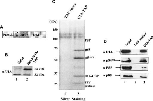
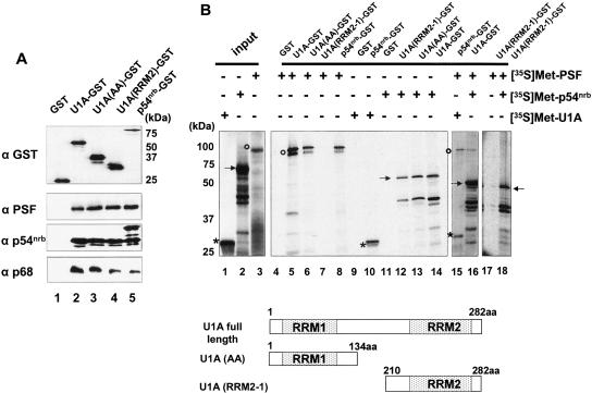
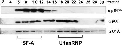
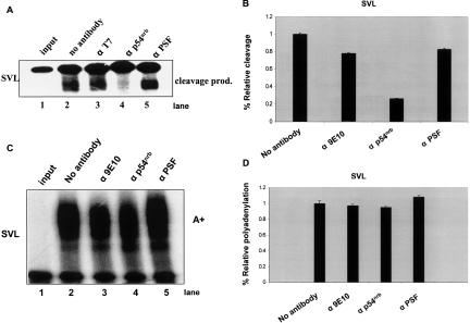
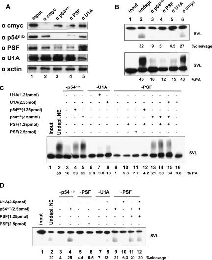
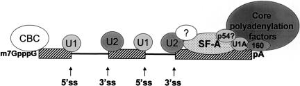
The U1 snRNP-A (U1A) protein has been known for many years as a component of the U1 snRNP. We have previously described a form of U1A present in human cells in significant amounts that is not associated with the U1 snRNP or U1 RNA but instead is part of a novel complex of non-snRNP proteins that we have termed snRNP-free U1A, or SF-A. Antibodies that specifically recognize this complex inhibit in vitro splicing and polyadenylation of pre-mRNA, suggesting that this complex may play an important functional role in these mRNA-processing activities. This finding was underscored by the determination that one of the components of this complex is the polypyrimidine-tract-binding protein-associated splicing factor, PSF. In order to further our studies on this complex and to determine the rest of the components of the SF-A complex, we prepared several stable HeLa cell lines that overexpress a tandem-affinity-purification-tagged version of U1A (TAP-tagged U1A). Nuclear extract was prepared from one of these cell lines, line 107, and affinity purification was performed along with RNase treatment. We have used mass spectrometry analysis to identify the candidate factors that associate with U1A. We have now identified and characterized PSF, p54(nrb), and p68 as novel components of the SF-A complex. We have explored the function of this complex in RNA processing, specifically cleavage and polyadenylation, by performing immunodepletions followed by reconstitution experiments, and have found that p54(nrb) is critical.


Figures

FIGURE 1.
FIGURE 1.
Purification of U1A interacting proteins. (A) Schematic representation of the U1A-TAP construct. U1A cDNA was cloned into pZome-1N (Cellzome) so that an N-terminal TAP cassette was introduced in frame with the U1A cDNA. The TAP tag consisted of a calmodulin-binding peptide (CBP), a consensus TEV cleavage site (TEV), and the IgG-binding domain of protein A (Prot.A). (B) The U1A-TAP construct was transfected into HeLa cells; stable, clonally isolated cell lines were established; and aliquots of nuclear proteins from these stable cell lines were analyzed by Western blot analysis with the U1A-specific polyclonal antibody 310. Note that the tagged U1A migrated with ~20 kDa greater apparent mass than endogenous U1A. (C) Analysis of U1A interacting proteins. Proteins of TAP elutes from either TAPvector-transfected or U1A-TAP-transfected cells were resolved on a 12.5% SDS-PAGE gel and silver stained. The proteins in lane 2 were identified by mass spectrometry as indicated on the right. The nuclear extract used in this TAP purification is pretreated with RNase A (see Materials and Methods). The protein band at ~27 kDa is the recombinant TEV protease. (D) Verification of the identities of the proteins from TAP-U1A purification. Aliquots of TEV eluates were subjected to Western blot analysis with specific antibodies against p54nrb, PSF, and p68, respectively. Lane 1 in each panel represents 10% of input.
FIGURE 2.
FIGURE 2.
Direct protein–protein interactions. (A) GST pull-down experiments with GST-tagged proteins and HeLa nuclear extract. Bacterially expressed GST, GST-U1A, GST-U1A (AA), GST-U1A (RRM2-1), or GST-p54nrb proteins (see Materials and Methods) individually were immobilized on glutathione-Sepharose beads and after purification were analyzed by 12.5% SDS-PAGE and Western blotting using an anti-GST antibody (top panel). These purified proteins were then incubated with HeLa nuclear extract. After extensive washing, proteins bound to the beads were eluted in protein sample buffer, resolved on 12.5% SDS-PAGE gels, and were analyzed by Western blotting. The antibodies used in the Western blot are indicated on the left. (B) GST pull-down experiments with 35S-labeled proteins. In vitro transcribed and translated [35S]Met-labeled U1A, p54nrb, and PSF (left) were tested for binding to GST, GST-U1A, GST-U1A (AA), GST-U1A (RRM2-1), and GST-p54nrb fusion proteins. PSF is indicated by open circles; p54nrb, by solid arrows; and U1A, by asterisks. A schematic representation of the structure of U1A and the two truncated mutants, U1A (AA) and U1A (RRM2-1), is shown below.
FIGURE 3.
FIGURE 3.
Sucrose-gradient fractionation shows that p54nrb, p68, and U1A co-migrate in the SF-A-containing fractions. Sucrose gradients (5%–30%) were prepared using human 293T cell nucleoplasm. One-milliliter (1 mL) fractions were taken and 30 μL of every other fraction were separated by 12.5% SDS-PAGE and were blotted onto nitrocellulose. Three identical blots were probed with different antibodies: p54nrb, U1A 310, or p68 antibody. Smaller numbers represent fractions from the top of the gradient. The relative migration positions of the SF-A complex and the U1 snRNP are noted below.
FIGURE 4.
FIGURE 4.
p54nrb and PSF are involved in in vitro polyadenylation. (A) Both p54nrb and PSF antibodies inhibited in vitro polyadenylation with SVL. Cleavage/polyadenylation reactions (see Materials and Methods) with SVL were performed in the presence or the absence of each of the antibodies as shown on the top of each lane. The reaction products were resolved on a 5% polyacrylamide gel containing 8 M urea and visualized by PhosphorImager analysis. Anti-c-myc-tag epitope antibody (9E10) was included as a control. (B) Quantitation of A. The same in vitro cleavage/polyadenylation reactions as panel B were repeated in three independent experiments on both SVL (dark bars) and L3 (gray bars). Polyadenylation efficiency was quantitated by PhosphorImager analysis and ImageQuant software. Percent relative polyadenylation was represented by the polyadenylation efficiency in the presence of antibodies vs. the total input RNA. Error bars represent SD. (C) Cleavage/polyadenylation reactions on SVL substrate RNAs were performed in the presence of the rabbit pre-bleed (pre) or anti-PSF antisera.
FIGURE 5.
FIGURE 5.
p54nrb antibody inhibits in vitro cleavage on SVL. (A) In vitro cleavage reactions (see Materials and Methods). The cleavage reactions were performed in the presence or the absence of antibodies to p54nrb or PSF, respectively. T7 antibody represents anti-T7 gene antibody (Novagen) (Lutz-Freyermuth et al. 1990). Cleavage products are indicated on the right. (B) Cleavage efficiency was quantitated by PhosphorImager analysis and ImageQuant software and represented the same way as relative polyadenylation. Standard deviation was calculated from three independent experiments. (C) p54nrb and PSF antibodies did not show significant inhibition on in vitro polyadenylation with precleaved SVL. 32P-labeled SVL RNA was in vitro transcribed from HpaI-linearized pSP65-SVL, and similar polyadenylation reactions were performed as shown in Figure 4B ▶. (D) The polyadenylation efficiency was quantitated by the polyadenylation efficiency in the presence of antibody vs. the absence of antibody. Error bars represent SD.
FIGURE 6.
FIGURE 6.
Immunodepletion and reconstitution experiments reveal a critical role for p54nrb in polyadenylation. (A) Western blot of immunodepleted extracts. The specific proteins that were immunodepleted in each case are listed at the top of the figure; the antibodies used for Western blotting are shown to the left. Ten micrograms (10 μg) of nuclear extract was loaded in each lane. (B) Immunodepleted extracts were tested for their activity in cleavage alone (top) or cleavage/ polyadenylation (bottom). The percentages shown below are quantification of percent cleavage (top) or percent polyadenylation (bottom) of this particular experiment; upon three independent repeats of this experiment very similar results were obtained. (C) Reconstitution experiments of purified recombinant protein(s) to immunodepleted extracts, then used in in vitro polyadenylation reactions with SVL substrate RNA. (Lane 1) Input SVL RNA; (lane 2) regular, nondepleted nuclear extract; (lanes 35) p54nrb-depleted extract; (lanes 4,5) 1.25 and 2.5 pmol of purified GST-p54nrb additions, respectively; (lanes 68) U1A-depleted extract; (lanes 7,8) 1.25 and 2.5 pmol of purified GST-U1A additions, respectively; (lanes 916) PSF-depleted extracts; (lanes 10,11) 1.25 and 2.5 pmol of purified His-PSF additions, respectively; (lane 12) 1.25 pmol of His-PSF + 1.25 pmol of GST-p54nrb reconstitutions; (lane 13) 2.5 pmol of GST-p54nrb added back; (lane 14) 1.25 pmol of His-PSF + 2.5 pmol of GST-p54nrb reconstitutions; (lane 15) 1.25 pmol of His-PSF + 2.5 pmol of GST-p54nrb + 2.5 pmol of GST-U1A reconstitutions; (lane 16) 2.5 pmol of GST-U1A reconstitution. (D) Reconstitution experiments of purified recombinant protein(s) to immunodepleted extracts, then used in in vitro cleavage reactions with SVL substrate RNA. (Lane 1) Input SVL RNA; (lane 2) regular nuclear extract; (lanes 3,4) p54nrb-depleted nuclear extract (lane 4 also includes 2.5 pmol of GST-p54nrb reconstitutions); (lanes 5,6) PSF-depleted nuclear extract (lane 6 also includes 2.5 pmol of purified His-PSF); (lanes 7,8) U1A-depleted nuclear extract (lane 8 also includes 2.5 pmol of purified GST-U1A); (lanes 912) PSF-depleted nuclear extract (lane 10 also includes purified 2.5 pmol of GST-U1A, lane 11 also includes 1.25 pmol of His- PSF + 2.5 pmol of GST-p54nrb reconstitutions, lane 12 also includes 1.25 pmol of His-PSF + 2.5 pmol of p54nrb + 2.5 pmol of GST-U1A reconstitutions).
FIGURE 7.
FIGURE 7.
Model. The SF-A complex may be an adaptor between splicing and core polyadenylation factors. Not pictured for simplicity: SR proteins, U2AF, ASF/SF2. (5′ss) 5′-Splice site; (3′ss) 3′-splice site; (pA) polyadenylation signal; (U1 and U2) U1 and U2 snRNPs, respectively; lines indicate introns of a representative mammalian gene; striped boxes indicate exons of a representative mammalian gene.

References

    1. Allain, F.H.T., Howe, P.W.A., Neuhaus, D., and Varani, G. 1997. Structural basis for the RNA-binding specificity of human U1A protein. EMBO J. 16: 5764–5774. - PMC - PubMed
    1. Berget, S.M. 1995. Exon recognition in vertebrate splicing. J. Biol. Chem. 270: 2411–2414. - PubMed
    1. Bienroth, S., Keller, W., and Wahle, E. 1993. Assembly of a processive messenger RNA polyadenylation complex. EMBO J. 12: 585–594. - PMC - PubMed
    1. Black, D.L. 1995. Finding splice sites within a wilderness of RNA. RNA 1: 763–771. - PMC - PubMed
    1. Colgan, D. and Manley, J.L. 1997. Mechanism and regulation of mRNA polyadenylation. Genes & Dev. 11: 2755–2766. - PubMed

Publication types

MeSH terms

LinkOut - more resources