. 2006 Feb;7(2):212-8.
doi: 10.1038/sj.embor.7400593.

The Saccharomyces cerevisiae Sae2 protein negatively regulates DNA damage checkpoint signalling

Affiliations

The Saccharomyces cerevisiae Sae2 protein negatively regulates DNA damage checkpoint signalling

Michela Clerici et al. EMBO Rep. 2006 Feb.

Abstract

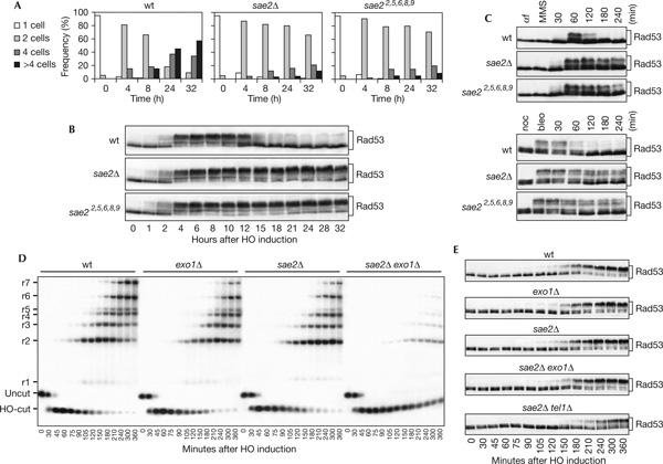
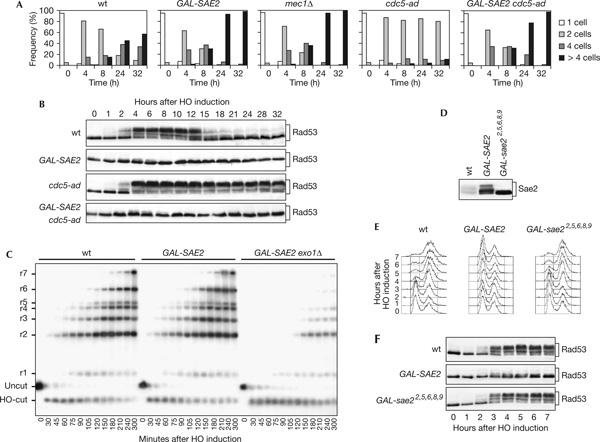
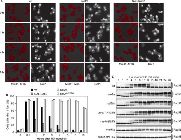
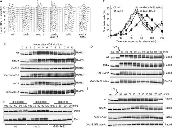
Double-strand breaks (DSBs) elicit a DNA damage response, resulting in checkpoint-mediated cell-cycle delay and DNA repair. The Saccharomyces cerevisiae Sae2 protein is known to act together with the MRX complex in meiotic DSB processing, as well as in DNA damage response during the mitotic cell cycle. Here, we report that cells lacking Sae2 fail to turn off both Mec1- and Tel1-dependent checkpoints activated by a single irreparable DSB, and delay Mre11 foci disassembly at DNA breaks, indicating that Sae2 may negatively regulate checkpoint signalling by modulating MRX association at damaged DNA. Consistently, high levels of Sae2 prevent checkpoint activation and impair MRX foci formation in response to unrepaired DSBs. Mec1- and Tel1-dependent Sae2 phosphorylation is necessary for these Sae2 functions, suggesting that the two kinases, once activated, may regulate checkpoint switch off through Sae2-mediated inhibition of MRX signalling.


Figures

Figure 1
Figure 1
Response to a single irreparable double-strand break in sae2Δ cells. (A) YEP+raf G1-arrested cell cultures of wild-type (wt) JKM139 and isogenic sae2Δ and sae22,5,6,8,9 strains were spotted on galactose-containing plates incubated at 30°C (time zero). At the indicated time points, 200 cells for each strain were analysed to determine the frequency of single cells and of cells forming microcolonies of two, four or more than four cells. (B) Galactose was added at time zero to wild-type JKM139 and isogenic sae2Δ and sae22,5,6,8,9 cell cultures exponentially growing in YEP+raf. Protein extracts from aliquots withdrawn at the indicated times were analysed by western blot with anti-Rad53 antibodies. (C) Wild-type W303 and isogenic sae2Δ and sae22,5,6,8,9 cell cultures arrested in G1 with α-factor (αf) or in G2 with nocodazole (noc) were incubated for 15 min with methylmethane sulphonate (MMS, 0.02%) or bleomycin (bleo, 10 mU/ml), respectively, and then released in YEPD. Protein extracts from samples taken at the indicated times were analysed by western blot with anti-Rad53 antibodies. (D) YEP+raf nocodazole-arrested cell cultures of wild-type JKM139 and isogenic exo1Δ, sae2Δ, sae2Δ exo1Δ and sae2Δ tel1Δ strains were transferred to YEP+raf+gal in the presence of nocodazole at time zero. Genomic DNA prepared from aliquots taken at the indicated times was digested with SspI and separated on alkaline agarose gel. Gel blots were hybridized with a single-stranded RNA probe specific for the MAT locus, which shows HO-cut and uncut fragments of 0.9 and 1.1 kb, respectively. As depicted in supplementary Fig S2 online, 5′-to-3′ resection progressively eliminates SspI sites located 1.7, 3.5, 4.7, 5.9, 6.5, 8.9 and 15.8 kb centromere-distal from the HO-cut site, producing larger SspI fragments (r1–r7) detected by the probe. The kinetics of resection product accumulation in sae2Δ tel1Δ cell cultures (not shown) was undistinguishable from that of sae2Δ cells. (E) Protein extracts from samples taken at the indicated times during the experiment in (D) were analysed by western blot with anti-Rad53 antibodies.
Figure 2
Figure 2
Response to a single irreparable double-strand break in SAE2-overexpressing cells. (A) YEP+raf G1-arrested cells of wild-type (wt) JKM139 and isogenic GAL-SAE2, cdc5-ad, GAL-SAE2 cdc5-ad and mec1Δ sml1Δ strains were spotted on galactose-containing plates incubated at 30°C (time zero). At the indicated times, 200 cells for each strain were analysed as in Fig 1A. (B) Galactose was added at time zero to wild-type JKM139 and isogenic GAL-SAE2, cdc5-ad and GAL-SAE2 cdc5-ad cell cultures exponentially growing in YEP+raf. Protein extracts from samples taken at the indicated times were subjected to western blot analysis with anti-Rad53 antibodies. (C) YEP+raf nocodazole-arrested cell cultures of wild-type JKM139 and isogenic GAL-SAE2 and GAL-SAE2 exo1Δ strains were transferred to YEP+raf+gal in the presence of nocodazole at time zero. Genomic DNA from samples collected at the indicated times was analysed as described in Fig 1D. (D) Cell cultures of JKM139 derivative strains carrying either the SAE2-HA allele at the SAE2 chromosomal locus or the GAL-SAE2-HA and GAL-sae22,5,6,8,9-HA fusions at the URA3 locus were shifted to YEP+raf+gal for 1 h and protein extracts were analysed by western blot with anti-haemagglutinin antibodies. (E,F) Exponentially growing YEP+raf cell cultures of wild-type JKM139 and isogenic GAL-SAE2 and GAL-sae22,5,6,8,9 strains were transferred to YEP+raf+gal at time zero. Aliquots were taken at the indicated times for fluorescence-activated cell sorting analysis (E) or western analysis with anti-Rad53 antibodies (F).
Figure 3
Figure 3
Mre11 localization at HO breaks in the absence or excess of Sae2. YEP+raf nocodazole-arrested cell cultures of wild-type (wt) JKM139 and isogenic sae2Δ and GAL-SAE2 strains, all carrying an MRE11-MYC-tagged allele at the MRE11 locus, were transferred to YEP+raf+gal in the presence of nocodazole (time zero). Cell samples taken at the indicated times were processed for staining with 4,6-diamidino-2-phenylindole (DAPI) and anti-Myc antibody indirect immunofluorescence. (A) Representative fields were photographed at the indicated times. (B) Kinetics of Mre11 foci formation were determined by scoring 200 cells for each strain at each time points. (C) Wild-type JKM139 and isogenic sae2Δ, rad50s, mre11-H125N, mre11-D56N, mre11Δ and mre11Δ sae2Δ strains, exponentially growing in YEP+raf, were transferred to YEP+raf+gal. Protein extracts from samples taken at the indicated times were analysed by western blot with anti-Rad53 antibodies.
Figure 4
Figure 4
Mec1- and Tel1-dependent checkpoints in sae2Δ and SAE2-overexpressing cells. (A,B) Exponentially growing YEP+raf cell cultures of wild-type YMV80 and isogenic sae2Δ, mec1Δ sml1Δ, sae2Δ mec1Δ sml1Δ, tel1Δ and sae2Δ tel1Δ strains were transferred to YEP+raf+gal to induce HO expression (time zero). Samples withdrawn at the indicated times were used for fluorescence-activated cell sorting analysis of DNA contents (A) and western blot analysis of protein extracts with anti-Rad53 antibodies (B). (C,D) Exponentially growing YEP+raf cell cultures of wild-type W303 and isogenic tel1Δ, GAL-SAE2 and GAL-SAE2 tel1Δ strains were arrested with nocodazole in the presence of galactose for 2 h (noc), UV-irradiated (45 J/m2) and released into cell cycle in YEP+raf+gal. Samples were collected at the indicated times to determine the percentage of binucleate cells in unirradiated (open symbols) and UV-irradiated (closed symbols) cultures (C) and to monitor Rad53 phosphorylation from UV-irradiated cell cultures by western analysis (D). (E) Exponentially growing YEP+raf cell cultures of wild-type W303 and isogenic mec1Δ sml1Δ, GAL-SAE2 and GAL-SAE2 mec1Δ sml1Δ strains were arrested in G1 with α factor in the presence of galactose for 2 h (αf), UV-irradiated (30 J/m2) and released into cell cycle in YEP+raf+gal. Protein extracts prepared at the indicated times were analysed by western blot with anti-Rad53 antibodies. (F) YEP+raf nocodazole-arrested cell cultures of wild-type W303 and isogenic sae2Δ and GAL-SAE2 strains, all expressing the DDC2-HA3- or MRE11-HA3-tagged alleles from the corresponding endogenous promoters, were transferred in YEP+raf+gal containing 10 mU/ml bleomycin and 15 μg/ml nocodazole (+bleo+noc). Protein extracts prepared at the indicated times were subjected to western analysis with anti-haemagglutinin antibodies.

References

    1. Baroni E, Viscardi V, Cartagena-Lirola H, Lucchini G, Longhese MP (2004) The functions of budding yeast Sae2 in the DNA damage response require Mec1- and Tel1-dependent phosphorylation. Mol Cell Biol 24: 4151–4165 - PMC - PubMed
    1. Clerici M, Mantiero D, Lucchini G, Longhese MP (2005) The Saccharomyces cerevisiae Sae2 protein promotes resection and bridging of double-strand break ends. J Biol Chem 280: 38631–38638 - PubMed
    1. Ira G et al. (2004) DNA end resection, homologous recombination and DNA damage checkpoint activation require CDK1. Nature 431: 1011–1017 - PMC - PubMed
    1. Keeney S, Kleckner N (1995) Covalent protein–DNA complexes at the 5′ strand termini of meiosis-specific double-strand breaks in yeast. Proc Natl Acad Sci USA 92: 11274–11278 - PMC - PubMed
    1. Lee SE, Moore JK, Holmes A, Umezu K, Kolodner RD, Haber JE (1998) Saccharomyces Ku70, Mre11/Rad50 and RPA proteins regulate adaptation to G2/M arrest after DNA damage. Cell 94: 399–409 - PubMed

Publication types

MeSH terms