. 2006 Jan;116(1):228-36.
doi: 10.1172/JCI25423. Epub 2005 Dec 22.

Neuronal eotaxin and the effects of CCR3 antagonist on airway hyperreactivity and M2 receptor dysfunction

Affiliations

Neuronal eotaxin and the effects of CCR3 antagonist on airway hyperreactivity and M2 receptor dysfunction

Allison D Fryer et al. J Clin Invest. 2006 Jan.

Abstract

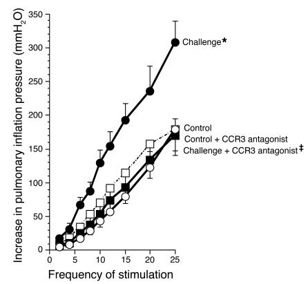
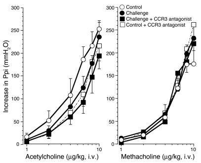
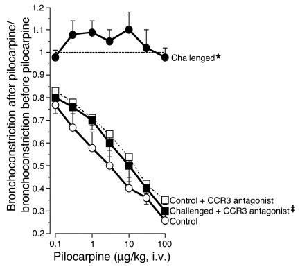
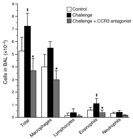
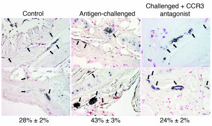
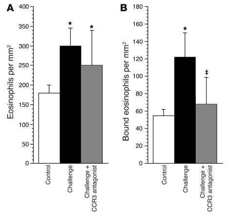
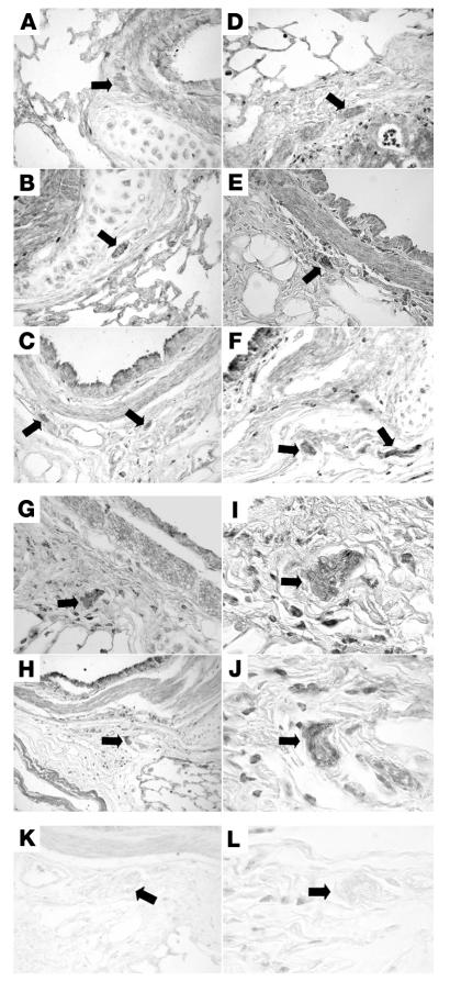
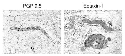
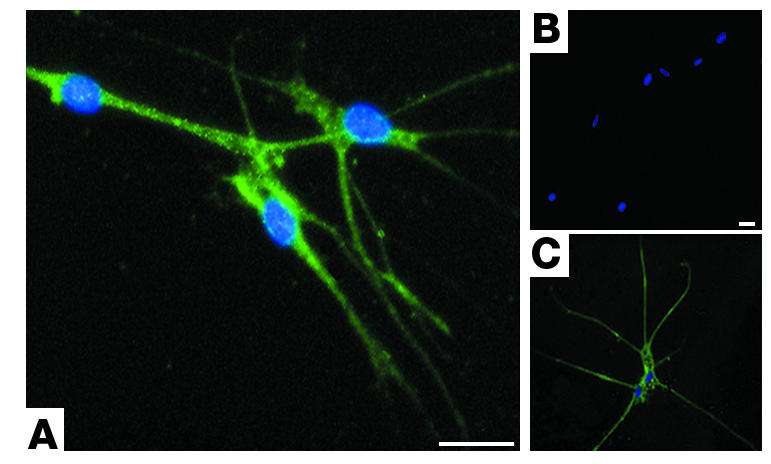
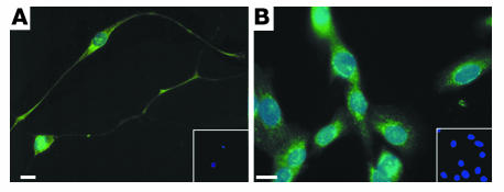
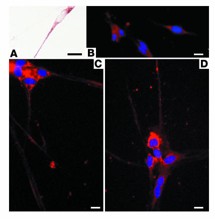
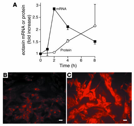
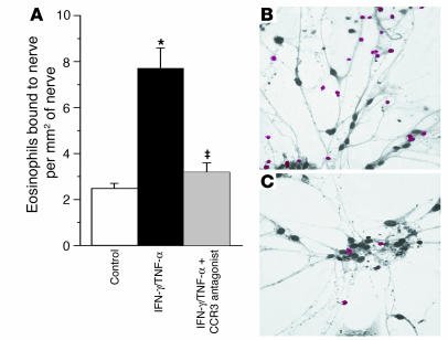
Eosinophils cluster around airway nerves in patients with fatal asthma and in antigen-challenged animals. Activated eosinophils release major basic protein, which blocks inhibitory M2 muscarinic receptors (M2Rs) on nerves, increasing acetylcholine release and potentiating vagally mediated bronchoconstriction. We tested whether GW701897B, an antagonist of CCR3 (the receptor for eotaxin as well as a group of eosinophil active chemokines), affected vagal reactivity and M2R function in ovalbumin-challenged guinea pigs. Sensitized animals were treated with the CCR3 antagonist before inhaling ovalbumin. Antigen-challenged animals were hyperresponsive to vagal stimulation, but those that received the CCR3 antagonist were not. M2R function was lost in antigen-challenged animals, but not in those that received the CCR3 antagonist. Although the CCR3 antagonist did not decrease the number of eosinophils in lung tissues as assessed histologically, CCR3 antagonist prevented antigen-induced clustering of eosinophils along the nerves. Immunostaining revealed eotaxin in airway nerves and in cultured airway parasympathetic neurons from both guinea pigs and humans. Both IL-4 and IL-13 increased expression of eotaxin in cultured airway parasympathetic neurons as well as in human neuroblastoma cells. Thus, signaling via CCR3 mediates eosinophil recruitment to airway nerves and may be a prerequisite to blockade of inhibitory M2Rs by eosinophil major basic protein.


Figures

Figure 1
Figure 1
Vagally mediated bronchoconstriction in control and antigen-challenged animals. Antigen challenge induced potentiation of vagally induced bronchoconstriction is inhibited by the CCR3 antagonist. In control animals (open circles), stimulation of both vagi (10 V, 0.2-millisecond pulse duration, 5-second pulse train) caused frequency-dependent bronchoconstriction that was significantly increased in antigen-challenged animals (filled circles). Pretreatment with the CCR3 antagonist (filled squares) prevented the increase in vagally induced bronchoconstriction in antigen-challenged guinea pigs. n = 5–7. *Significantly different from control, P = 0.001; significantly different from challenged, P = 0.0006. The CCR3 antagonist did not affect vagally induced bronchoconstriction in non–antigen-challenged control animals (open squares; n = 6).
Figure 2
Figure 2
Bronchoconstriction in response to exogenous cholinergic agonists. Bronchoconstriction induced by i.v. acetylcholine (left) or methacholine (right) in vagotomized animals was not changed by antigen challenge (filled circles) or by treatment of antigen-challenged animals with the CCR3 antagonist (filled squares). n = 5–7. The CCR3 antagonist did not affect bronchoconstriction in non–antigen-challenged control animals (open squares; n = 5). Ppi, pulmonary inflation pressure.
Figure 3
Figure 3
CCR3 antagonist prevents antigen-induced M2R dysfunction. Pilocarpine inhibited vagally induced bronchoconstriction in control animals (open circles) demonstrating functional M2Rs. Pilocarpine did not inhibit vagally induced bronchoconstriction in antigen-challenged animals (filled circles), demonstrating that the M2Rs were no longer responding to agonist stimulation. M2R dysfunction was prevented by the CCR3 antagonist (filled squares). n = 5–7. *Significantly different from control, P = 0.0006; significantly different from challenged, P = 0.001. The CCR3 antagonist did not affect the response to pilocarpine in non–antigen-challenged control animals (open squares; n = 5).
Figure 4
Figure 4
Inflammatory cells in lung lavage fluid. Antigen challenge caused a nonsignificant increase in total leukocytes, which consisted primarily of macrophages and eosinophils. This was prevented by the CCR3 antagonist. n = 5–7. Significantly different from control, P < 0.05; *significantly different from challenged, P < 0.05. BAL, bronchoalveolar lavage.
Figure 5
Figure 5
Treatment with a CCR3 antagonist prevents the increase in eosinophils around airway nerves after antigen challenge. Airway nerves (arrows) were stained black with antibody against PGP 9.5, and eosinophils were stained red with chromotrope 2R in control and antigen-challenged animals and in antigen-challenged animals treated with CCR3 antagonist. Numbers under the panels refer to the percentage of eosinophils (mean ± SEM; n = 5–7) in contact with airway nerves. The scale bar of 50 μm applies to all panels except the one in the upper right, where it equals 20 μm.
Figure 6
Figure 6
Antigen challenge increases eosinophils in airway tissues in general (A) as well as specifically in the area of airway nerves (B). The CCR3 antagonist did not prevent the general increase in airway tissue eosinophils (A). In contrast, the CCR3 antagonist selectively prevented the antigen-induced increase in eosinophils around the nerves (B). n = 5–7. *Significantly different from control, P < 0.01; significantly different from challenged, P < 0.01.
Figure 7
Figure 7
Immunostaining of airway tissues for eotaxin. Eotaxin expression was present throughout the airways, including in the nerves (arrows), of control guinea pigs (AC) and was increased after antigen challenge (DJ). Staining of nerves (assessed as mean pixel intensity) was increased from 64.8 ± 5.9 in controls to 114.3 ± 7.8 in antigen-challenged animals (P = 0.002). Nerve bundles in antigen-challenged guinea pigs are shown under low (G and H) and high magnification (I and J). There was no positive eotaxin stain in lungs or nerves (arrows) of either control (K) or antigen-challenged (L) lung when preimmune serum was used in place of the antibody. Scale bars: 50 μm.
Figure 8
Figure 8
Immunostaining of human airway nerves for eotaxin. Airway nerves were labeled with an antibody against PGP 9.5 (a marker of nerves; left panel) and an adjacent section labeled with antibody against human eotaxin (right panel). The eotaxin antibody labeled nerves (N) and glands (G) in a nonasthmatic individual. Scale bar: 10 μm.
Figure 9
Figure 9
Primary cultures of human airway parasympathetic nerves express eotaxin. Neurons are stained with antibody against eotaxin (A), no primary antibody (B), or with antibody against the nerve-specific protein PGP 9.5 (C). All cells are stained with DAPI, labeling nuclei blue. Scale bars: 25 μM.
Figure 10
Figure 10
Immunostaining for IL-4 receptor. IL-4 receptors were present on primary cultures of guinea pig airway parasympathetic neurons (A) and on human neuroblastoma cells (B). Neurons were stained with antibody against IL-4 (green) and DAPI, which stains the nuclei blue. Insets show cells in the absence of primary antibody; only blue DAPI-stained nuclei are visible (10-fold lower magnification). Scale bars: 30 μm.
Figure 11
Figure 11
Primary cultures of guinea pig airway parasympathetic nerves stain positively with an antibody against eotaxin (AD). Faint staining in untreated cells (B) was increased by treatment with IL-4 (C and D) or IL-13 (data not shown). Scale bars: 30 μm.
Figure 12
Figure 12
IL-4 stimulates expression of eotaxin in human neuroblastoma cells. eotaxin mRNA and protein expression were increased after exposure to IL-4 (1 ng/ml) (A). Immunostaining demonstrated increased eotaxin protein 8 hours after exposure to IL-4 (C) compared with untreated controls (B). Scale bars: 30 μm.
Figure 13
Figure 13
CCR3 antagonist inhibits eosinophil adhesion to primary cultures of parasympathetic nerves. Eosinophil adhesion to nerve cells was significantly increased in cells treated for 24 hours with IFN-γ (1,000 U/ml) and TNF-α (2 ng/ml) (A, black bar), compared with controls (A, white bar). The CCR3 antagonist significantly inhibited adherence of eosinophils to the nerves (A, gray bar). Representative photos of eosinophil (red) adhesion to nerves treated with IFN-γ and TNF-α (IFN-γ/TNF-α) in the absence (B) or presence (C) of the CCR3 antagonist are shown. n = 4. *Significantly different from control, P = 0.01; significantly different from challenged, P = 0.04. Scale bar: 20 μm.

References

    1. Fryer AD, Maclagan J. Muscarinic inhibitory receptors in pulmonary parasympathetic nerves in the guinea-pig. Br. J. Pharmacol. 1984;83:973–978. - PMC - PubMed
    1. Minette P, Barnes PJ. Prejunctional inhibitory muscarinic receptors on cholinergic nerves in human and guinea-pig airways. J. Appl. Physiol. 1988;64:2532–2537. - PubMed
    1. Fryer AD, Jacoby DB. Function of pulmonary M2 muscarinic receptors in antigen challenged guinea-pigs is restored by heparin and poly-l-glutamate. J. Clin. Invest. 1992;90:2292–2298. - PMC - PubMed
    1. Minette PJ, Lammers JWJ, Dixon CMS, McCusker MT, Barnes PJ. A muscarinic agonist inhibits reflex bronchoconstriction in normal but not asthmatic subjects. J. Appl. Physiol. 1989;67:2461–2465. - PubMed
    1. Costello RW, et al. Localization of eosinophils to airway nerves and effect on neuronal M2 muscarinic receptor function. Am. J. Physiol. 1997;273:L93–L103. - PubMed

Publication types

MeSH terms