Pyramidal cells of the rat basolateral amygdala: synaptology and innervation by parvalbumin-immunoreactive interneurons
- PMID: 16374802
- PMCID: PMC2562221
- DOI: 10.1002/cne.20832
Pyramidal cells of the rat basolateral amygdala: synaptology and innervation by parvalbumin-immunoreactive interneurons
Abstract
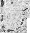
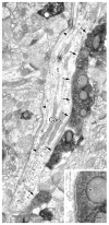
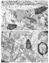
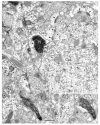
The generation of emotional responses by the basolateral amygdala is determined largely by the balance of excitatory and inhibitory inputs to its principal neurons, the pyramidal cells. The activity of these neurons is tightly controlled by gamma-aminobutyric acid (GABA)-ergic interneurons, especially a parvalbumin-positive (PV(+)) subpopulation that constitutes almost half of all interneurons in the basolateral amygdala. In the present semiquantitative investigation, we studied the incidence of synaptic inputs of PV(+) axon terminals onto pyramidal neurons in the rat basolateral nucleus (BLa). Pyramidal cells were identified by using calcium/calmodulin-dependent protein kinase II (CaMK) immunoreactivity as a marker. To appreciate the relative abundance of PV(+) inputs compared with excitatory inputs and other non-PV(+) inhibitory inputs, we also analyzed the proportions of asymmetrical (presumed excitatory) synapses and symmetrical (presumed inhibitory) synapses formed by unlabeled axon terminals targeting pyramidal neurons. The results indicate that the perisomatic region of pyramidal cells is innervated almost entirely by symmetrical synapses, whereas the density of asymmetrical synapses increases as one proceeds from thicker proximal dendritic shafts to thinner distal dendritic shafts. The great majority of synapses with dendritic spines are asymmetrical. PV(+) axon terminals form mainly symmetrical synapses. These PV(+) synapses constitute slightly more than half of the symmetrical synapses formed with each postsynaptic compartment of BLa pyramidal cells. These data indicate that the synaptology of basolateral amygdalar pyramidal cells is remarkably similar to that of cortical pyramidal cells and that PV(+) interneurons provide a robust inhibition of both the perisomatic and the distal dendritic domains of these principal neurons.
J. Comp. Neurol. 494:635-650, 2006. (c) 2005 Wiley-Liss, Inc.
Figures


Similar articles
-
Postsynaptic targets of somatostatin-containing interneurons in the rat basolateral amygdala.J Comp Neurol. 2007 Jan 20;500(3):513-29. doi: 10.1002/cne.21185. J Comp Neurol. 2007. PMID: 17120289
-
Cholinergic innervation of pyramidal cells and parvalbumin-immunoreactive interneurons in the rat basolateral amygdala.J Comp Neurol. 2011 Mar 1;519(4):790-805. doi: 10.1002/cne.22550. J Comp Neurol. 2011. PMID: 21246555 Free PMC article.
-
GABAergic innervation of alpha type II calcium/calmodulin-dependent protein kinase immunoreactive pyramidal neurons in the rat basolateral amygdala.J Comp Neurol. 2002 May 6;446(3):199-218. doi: 10.1002/cne.10204. J Comp Neurol. 2002. PMID: 11932937
-
Coupled networks of parvalbumin-immunoreactive interneurons in the rat basolateral amygdala.J Neurosci. 2005 Aug 10;25(32):7366-76. doi: 10.1523/JNEUROSCI.0899-05.2005. J Neurosci. 2005. PMID: 16093387 Free PMC article.
-
Dopaminergic innervation of pyramidal cells in the rat basolateral amygdala.Brain Struct Funct. 2009 Feb;213(3):275-88. doi: 10.1007/s00429-008-0196-y. Epub 2008 Oct 7. Brain Struct Funct. 2009. PMID: 18839210
Cited by
-
Repeated restraint stress increases basolateral amygdala neuronal activity in an age-dependent manner.Neuroscience. 2012 Dec 13;226:459-74. doi: 10.1016/j.neuroscience.2012.08.051. Epub 2012 Sep 15. Neuroscience. 2012. PMID: 22986163 Free PMC article.
-
Mild Traumatic Brain Injury Produces Neuron Loss That Can Be Rescued by Modulating Microglial Activation Using a CB2 Receptor Inverse Agonist.Front Neurosci. 2016 Oct 6;10:449. doi: 10.3389/fnins.2016.00449. eCollection 2016. Front Neurosci. 2016. PMID: 27766068 Free PMC article.
-
Inhibitory Circuits in the Basolateral Amygdala in Aversive Learning and Memory.Front Neural Circuits. 2021 Apr 30;15:633235. doi: 10.3389/fncir.2021.633235. eCollection 2021. Front Neural Circuits. 2021. PMID: 33994955 Free PMC article. Review.
-
Amygdala microcircuits controlling learned fear.Neuron. 2014 Jun 4;82(5):966-80. doi: 10.1016/j.neuron.2014.04.042. Neuron. 2014. PMID: 24908482 Free PMC article. Review.
-
Hemispheric differences in the number of parvalbumin-positive neurons in subdivisions of the rat basolateral amygdala complex.Brain Res. 2018 Jan 1;1678:214-219. doi: 10.1016/j.brainres.2017.10.028. Epub 2017 Oct 28. Brain Res. 2018. PMID: 29107660 Free PMC article.
References
-
- Aggleton JP. The Amygdala: Neurobiological Aspects of Emotion, Memory, and Mental Dysfunction. Wiley-Liss; New York: 1992.
-
- Aggleton JP. The Amygdala: A Functional Analysis. Wiley-Liss; New York: 2000.
-
- Alonso-Nanclares L, White EL, Elston GN, DeFelipe J. Synaptology of the proximal segment of pyramidal cell basal dendrites. Eur J Neurosci. 2004;19:771–6. - PubMed
-
- Asan E. The catecholaminergic innervation of the rat amygdala. Adv Anat Embryol Cell Biol. 1998;142:1–118. - PubMed
-
- Acsady L, Arabadzisz D, Freund TF. Correlated morphological and neurochemical features identify different subsets of vasoactive intestinal polypeptide-immunoreactive interneurons in rat hippocampus. Neuroscience. 1996;73:299–315. - PubMed
Publication types
MeSH terms
Substances
Grants and funding
LinkOut - more resources
Full Text Sources

