

Afferents to the orexin neurons of the rat brain
- PMID: 16374809
- PMCID: PMC2259441
- DOI: 10.1002/cne.20859
Afferents to the orexin neurons of the rat brain
Abstract
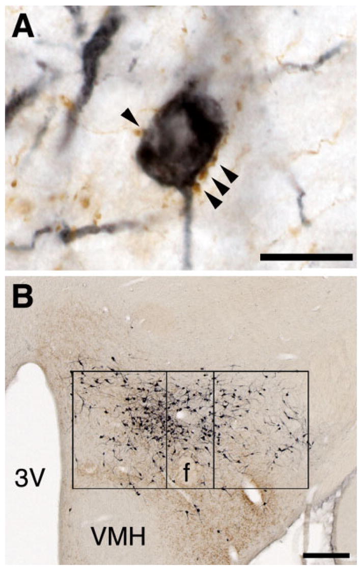
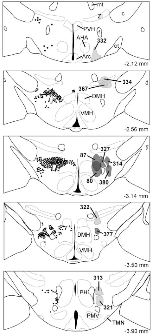
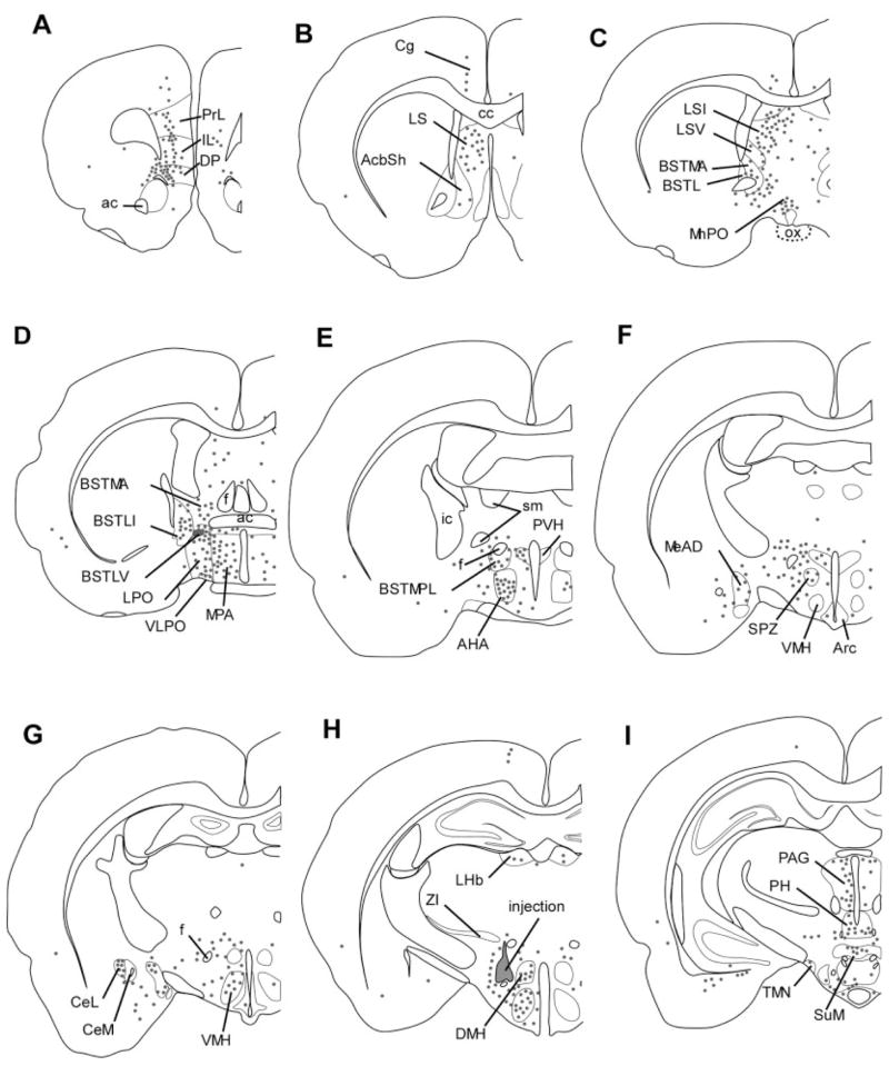
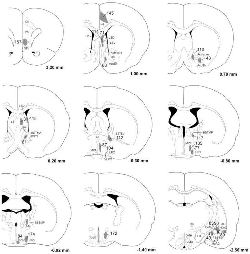
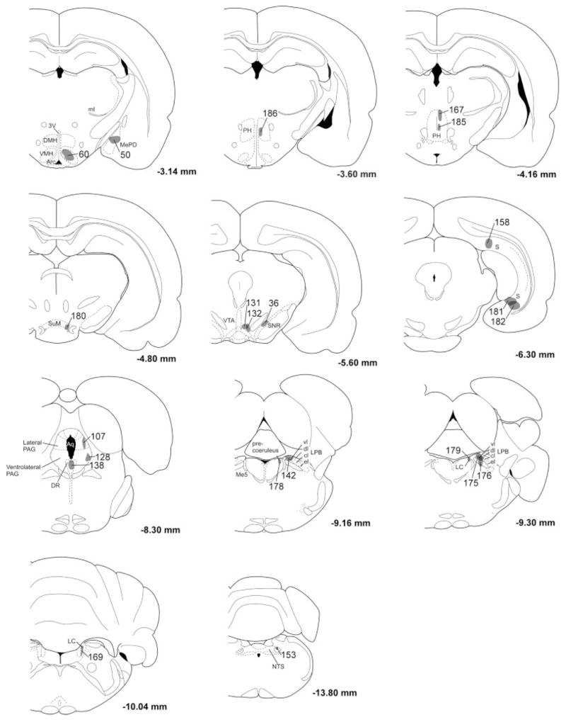
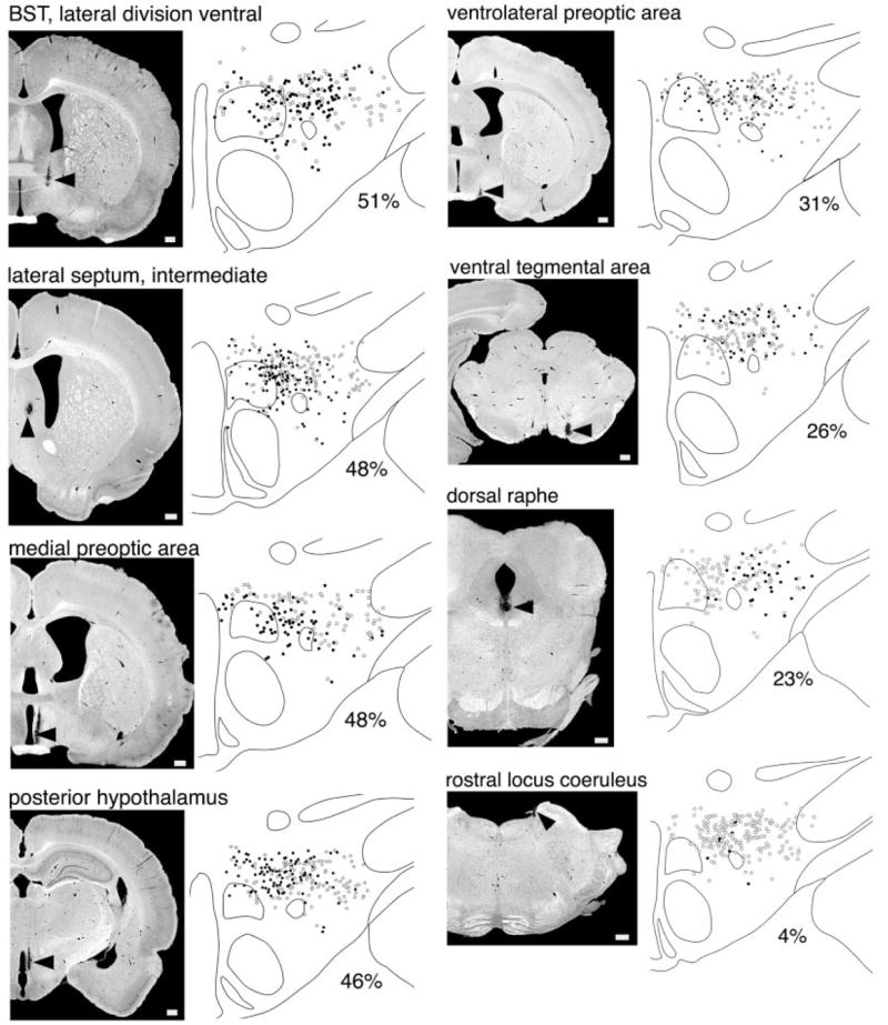
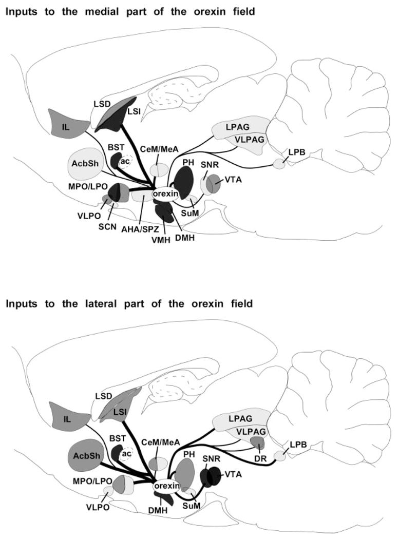
Emotions, stress, hunger, and circadian rhythms all promote wakefulness and behavioral arousal. Little is known about the pathways mediating these influences, but the orexin-producing neurons of the hypothalamus may play an essential role. These cells heavily innervate many wake-promoting brain regions, and mice lacking the orexin neurons have narcolepsy and fail to rouse in response to hunger (Yamanaka et al. [2003] Neuron 38:701-713). To identify the afferents to the orexin neurons, we first injected a retrograde tracer into the orexin neuron field of rats. Retrogradely labeled neurons were abundant in the allocortex, claustrum, lateral septum, bed nucleus of the stria terminalis, and in many hypothalamic regions including the preoptic area, dorsomedial nucleus, lateral hypothalamus, and posterior hypothalamus. Retrograde labeling in the brainstem was generally more modest, but labeling was strong in the periaqueductal gray matter, dorsal raphe nucleus, and lateral parabrachial nucleus. Injection of an anterograde tracer confirmed that most of these regions directly innervate the orexin neurons, with some of the heaviest input coming from the lateral septum, preoptic area, and posterior hypothalamus. In addition, hypothalamic regions preferentially innervate orexin neurons in the medial and perifornical parts of the field, but most projections from the brainstem target the lateral part of the field. Inputs from the suprachiasmatic nucleus are mainly relayed via the subparaventricular zone and dorsomedial nucleus. These observations suggest that the orexin neurons may integrate a variety of interoceptive and homeostatic signals to increase behavioral arousal in response to hunger, stress, circadian signals, and autonomic challenges.
Copyright 2005 Wiley-Liss, Inc.
Figures

Similar articles
-
Retrograde study of hypocretin-1 (orexin-A) projections to subdivisions of the dorsal raphe nucleus in the rat.Brain Res. 2005 Oct 12;1059(1):35-45. doi: 10.1016/j.brainres.2005.08.016. Epub 2005 Sep 8. Brain Res. 2005. PMID: 16153616
-
Inputs to the ventrolateral bed nucleus of the stria terminalis.J Comp Neurol. 2008 Dec 10;511(5):628-57. doi: 10.1002/cne.21870. J Comp Neurol. 2008. PMID: 18853414 Free PMC article.
-
Indirect projections from the suprachiasmatic nucleus to major arousal-promoting cell groups in rat: implications for the circadian control of behavioural state.Neuroscience. 2005;130(1):165-83. doi: 10.1016/j.neuroscience.2004.08.030. Neuroscience. 2005. PMID: 15561433
-
[Hypothalamic neuropeptides implicated in the regulation of sleep/wakefulness states].Brain Nerve. 2012 Jun;64(6):629-37. Brain Nerve. 2012. PMID: 22647470 Review. Japanese.
-
Control of arousal by the orexin neurons.Curr Opin Neurobiol. 2013 Oct;23(5):752-9. doi: 10.1016/j.conb.2013.04.008. Epub 2013 May 15. Curr Opin Neurobiol. 2013. PMID: 23683477 Free PMC article. Review.
Cited by
-
Central control of circadian phase in arousal-promoting neurons.PLoS One. 2013 Jun 24;8(6):e67173. doi: 10.1371/journal.pone.0067173. Print 2013. PLoS One. 2013. PMID: 23826226 Free PMC article.
-
Neurotensin co-expressed in orexin-producing neurons in the lateral hypothalamus plays an important role in regulation of sleep/wakefulness states.PLoS One. 2013 Apr 19;8(4):e62391. doi: 10.1371/journal.pone.0062391. Print 2013. PLoS One. 2013. PMID: 23620827 Free PMC article.
-
Hypothalamic orexinergic neuron changes during the hibernation of the Syrian hamster.Front Neuroanat. 2022 Sep 9;16:993421. doi: 10.3389/fnana.2022.993421. eCollection 2022. Front Neuroanat. 2022. PMID: 36157325 Free PMC article.
-
Forebrain networks and the control of feeding by environmental learned cues.Physiol Behav. 2013 Sep 10;121:10-8. doi: 10.1016/j.physbeh.2013.03.024. Epub 2013 Apr 3. Physiol Behav. 2013. PMID: 23562305 Free PMC article. Review.
-
Role of the basal ganglia in the control of sleep and wakefulness.Curr Opin Neurobiol. 2013 Oct;23(5):780-5. doi: 10.1016/j.conb.2013.02.001. Epub 2013 Apr 24. Curr Opin Neurobiol. 2013. PMID: 23465424 Free PMC article. Review.
References
-
- Alam N, Kumar S, Bashir T, Suntsova N, Szymusiak R, McGinty D. Muscimol microinjection into median preoptic nucleus (MnPN) activates c-fos in hypocretin and other perifornical-lateral hypothalamic area (PF-LHA) neurons. Annual Meeting of the Society for Neuroscience; San Diego. 2004.
-
- Alheid GF, de Olmos JS, Beltramino CA. Amygdala and extended amygdala. In: Paxinos G, editor. The rat nervous system. San Diego: Academic Press; 1995. pp. 495–578.
-
- Baldo BA, Gual-Bonilla L, Sijapati K, Daniel RA, Landry CF, Kelley AE. Activation of a subpopulation of orexin/hypocretin-containing hypothalamic neurons by GABAA receptor-mediated inhibition of the nucleus accumbens shell, but not by exposure to a novel environment. Eur J Neurosci. 2004;19:376–386. - PubMed
-
- Bonnet MH, Arand DL. Heart rate variability in insomniacs and matched normal sleepers. Psychosom Med. 1998;60:610–615. - PubMed
-
- Bonnet MH, Arand DL. Insomnia, metabolic rate and sleep restoration. J Intern Med. 2003;254:23–31. - PubMed
Publication types
MeSH terms
Substances
Grants and funding
LinkOut - more resources
Full Text Sources
Other Literature Sources
