. 2005 Dec;2(5):387-99.
doi: 10.2174/156720205774962683.

Erythropoietin requires NF-kappaB and its nuclear translocation to prevent early and late apoptotic neuronal injury during beta-amyloid toxicity

Affiliations

Erythropoietin requires NF-kappaB and its nuclear translocation to prevent early and late apoptotic neuronal injury during beta-amyloid toxicity

Zhao Zhong Chong et al. Curr Neurovasc Res. 2005 Dec.

Abstract

No longer considered exclusive for the function of the hematopoietic system, erythropoietin (EPO) is now considered as a viable agent to address central nervous system injury in a variety of cellular systems that involve neuronal, vascular, and inflammatory cells. Yet, it remains unclear whether the protective capacity of EPO may be effective for chronic neurodegenerative disorders such as Alzheimer's disease (AD) that involve beta-amyloid (Abeta) apoptotic injury to hippocampal neurons. We therefore investigated whether EPO could prevent both early and late apoptotic injury during Abeta exposure in primary hippocampal neurons and assessed potential cellular pathways responsible for this protection. Primary hippocampal neuronal injury was evaluated by trypan blue dye exclusion, DNA fragmentation, membrane phosphatidylserine (PS) exposure, and nuclear factor-kappaB (NF-kappaB) expression with subcellular translocation. We show that EPO, in a concentration specific manner, is able to prevent the loss of both apoptotic genomic DNA integrity and cellular membrane asymmetry during Abeta exposure. This blockade of Abeta generated neuronal apoptosis by EPO is both necessary and sufficient, since protection by EPO is completely abolished by co-treatment with an anti-EPO neutralizing antibody. Furthermore, neuroprotection by EPO is closely linked to the expression of NF-kappaB p65 by preventing the degradation of this protein by Abeta and fostering the subcellular translocation of NF-kappaB p65 from the cytoplasm to the nucleus to allow the initiation of an anti-apoptotic program. In addition, EPO intimately relies upon NF-kappaB p65 to promote neuronal survival, since gene silencing of NF-kappaB p65 by RNA interference removes the protective capacity of EPO during Abeta exposure. Our work illustrates that EPO is an effective entity at the neuronal cellular level against Abeta toxicity and requires the close modulation of the NF-kappaB p65 pathway, suggesting that either EPO or NF-kappaB may be used as future potential therapeutic strategies for the management of chronic neurodegenerative disorders, such as AD.


Figures
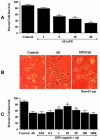
Fig. (1)
Fig. (1). Erythropoietin (EPO) increases neuronal survival during β-amyloid (Aβ) toxicity and is concentration specific
(A) Aβ was applied to primary hippocampal neuronal cultures at a concentration of 1, 5, 10, 20 μM and neuronal survival was determined 24 hours later by using the trypan blue exclusion method. Neuronal survival was progressively decreased with increased concentration of Aβ (*P<0.01 vs. untreated control). (B) Representative images illustrate trypan blue staining. Aβ (5 μM) applied to neuronal cultures resulted in significant staining 24 hours following Aβ treatment. EPO (10 ng/ml) applied 1 hour prior to Aβ (5 μM) exposure significantly increased neuronal survival and prevented trypan blue uptake 24 hours following Aβ treatment. (C) Neurons were pretreated with EPO (0.01 to 1000 ng/ml) 1 hour prior to Aβ (5 μM) exposure and cell survival was assessed 24 hours later. Protection of EPO against Aβ toxicity was evident in cultures with EPO (1 to 50 ng/ml) when compared with cultures exposed to Aβ alone (*P<0.01 vs. Aβ treated alone). In A and C, each data point represents the mean and SEM.
Fig. (2)
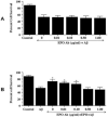
Fig. (2). Erythropoietin (EPO) is necessary and sufficient for neuronal protection against β-amyloid (Aβ) toxicity
(A) The EPO blocking antibody (EPO Ab) (0.01-1 μg/ml) was applied 1 hour prior to Aβ (5 μM) application. Neuronal survival was assessed 24 hours later. No significant changes in neuronal survival were observed following application of the EPO Ab when compared to cultures treated with Aβ alone. (B) Increasing concentrations of the EPO Ab (0.01 - 1.00 μg/ml) were applied to neuronal cultures in conjunction with EPO (10 ng/ml) for 1 hour prior to Aβ (5 μM) application. Neuronal survival was assessed 24 hours later. Protection by EPO against Aβ toxicity was attenuated or abolished during application of the EPO Ab (0.50 and 1.00 μg/ml) (*P<0.01 vs. Aβ treated alone). In A and B, each data point represents the mean and SEM.
Fig. (3)
Fig. (3). Erythropoietin (EPO) prevents apoptotic genomic DNA fragmentation and membrane phosphatidylserine (PS) exposure during β-amyloid (Aβ) application
(A) Neurons were pretreated with EPO (10 ng/ml) 1 hour prior to Aβ application. DNA fragmentation was assessed using TUNEL and membrane PS exposure was determined by annexin V phycoerythrin labeling 24 hours following Aβ treatment. Representative images illustrate DNA fragmentation and membrane PS externalization in neurons using transmitted (T) light and fluorescence (F) light of the same microscopy field with 490 nm excitation and 585 nm emission wavelengths. EPO significantly prevented DNA fragmentation and membrane PS exposure during Aβ exposure. (B) Pretreatment with EPO (10 ng/ml) 1 hour prior to Aβ application decreased DNA fragmentation and membrane PS externalization significantly 24 hours following Aβ exposure (*P<0.01 vs. Aβ). In B, each data point represents the mean and SEM, control = untreated neurons.
Fig. (4)
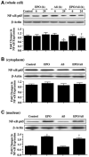
Fig. (4). Erythropoietin (EPO) prevents the degradation of NF-κ B and leads to its nuclear translocation during β-amyloid (Aβ) toxicity
(A) Equal amounts of neuronal protein extracts (50 μg/lane) were immunoblotted 6 and 24 hours following application of Aβ (5 μM), EPO (10 ng/ml), or with EPO (10 ng/ml) 1 hour pretreatment prior to Aβ with anti NF-κB p65 antibody. Exposure to Aβ decreased the expression NF-κB p65 at 24 hours following Aβ application, but EPO pretreatment prevented the decrease in the expression of NF-κB p65 during Aβ exposure (*P<0.01 vs. control; †P< 0.01 vs. Aβ 24 hours). (B) and (C) EPO (10 ng/ml) was applied 1 hour prior to Aβ (5 μM) administration and cellular cytoplasm and nuclear proteins were prepared separately at 6 hours following Aβ application. The expression of NF-κB p65 in cytoplasmic extracts (B) and nuclear extracts (C) was determined by Western blot analysis. No significant change in the expression of NF-κB p65 was observed in the cytoplasmic extracts (B). Yet, EPO lead to a significant increase in nuclear NF-B p65 expression either alone or during Aβ application (C) (*P<0.01 vs. control; †P< 0.01 vs. Aβ). In A, B, and C, each data point represents the mean and SEM, control = untreated cells.
Fig. (5)
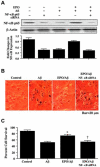
Fig. (5). Erythropoietin requires NF-κ B p65 to increase neuronal survival during exposure to β-amyloid (Aβ)
(A) Neurons were transfected with NF-κB p65 siRNA for 72 hours prior to EPO and Aβ application. Western blot analysis for NF-κB p65 was performed 6 hours following Aβ administration. EPO (10 ng/ml) applied 1 hour prior to Aβ (5 μM) prevented the degradation of NF-κB p65. In contrast, NF-κB p65 siRNA gene silencing significantly reduced the efficacy of EPO and decreased the expression of NF-κB p65 in neurons during untreated cultures and during application of combined EPO and Aβ. (B) Representative images illustrate that Aβ (5 μM) exposure leads to significant trypan blue uptake in neurons 24 hours following Aβ treatment. EPO (10 ng/ml) given 1 hour prior to Aβ application significantly reduced trypan blue uptake in neurons indicative of the maintenance of intact cellular membranes. In contrast, NF-κB p65 siRNA transfection for 72 hours prior to EPO application reduced the protective capacity of EPO resulting in neuronal injury with significant trypan blue uptake. (C) Neuronal survival is significantly increased by EPO (10 ng/ml) applied 1 hour prior to Aβ (5 μM) when compared to Aβ treated neurons alone. In contrast, protection conferred by EPO against Aβ toxicity is lost during NF-κB p65 gene silencing with targeted siRNA (*P<0.01 vs. Aβ treated alone; †P<0.01 vs. EPO/Aβ). Each data point represents the mean and SEM. Control = untreated cultures.
Fig. (6)
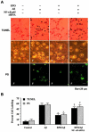
Fig. (6). Prevention of apoptotic genomic DNA fragmentation and cellular membrane PS exposure by EPO during β-amyloid (Aβ) toxicity is mediated by NF-κB p65
(A) Neurons were transfected with NF-κB p65 siRNA for 72 hours prior to EPO and Aβ application. DNA fragmentation was assessed 24 hours after Aβ administration using TUNEL and PS exposure was determined by annexin V phycoerythrin labeling 24 hours following Aβ administration. Representative images illustrate DNA fragmentation and membrane PS externalization in neurons using transmitted (T) light and fluorescence (F) light of the same microscopy field with 490 nm excitation and 585 nm emission wavelengths. EPO (10 ng/ml) applied 1 hour prior to Aβ administration significantly prevented DNA fragmentation and membrane PS exposure following Aβ. In contrast, gene silencing of NF-κB p65 with siRNA abrogates protection of EPO in neurons and yields a significant increase in positive TUNEL and cellular membrane PS externalization. (B) NF-κB p65 gene silencing with targeted siRNA eliminates protection conferred by EPO against Aβ, resulting in an increase in percent DNA fragmentation and cellular membrane PS exposure (*P<0.01 vs. Aβ treated alone; †P<0.01 vs. EPO/Aβ). Each data point represents the mean and SEM. Control = untreated cultures.

Similar articles

Cited by

References

    1. Bernaudin M, Marti HH, Roussel S, Divoux D, Nouvelot A, MacKenzie ET, Petit E. A potential role for erythropoietin in focal permanent cerebral ischemia in mice. J Cereb Blood Flow Metab. 1999;19(6):643–51. - PubMed
    1. Bittorf T, Buchse T, Sasse T, Jaster R, Brock J. Activation of the transcription factor NF-kappaB by the erythropoietin receptor: structural requirements and biological significance. Cell Signal. 2001;13(9):673–81. - PubMed
    1. Bornemann KD, Wiederhold KH, Pauli C, Ermini F, Stalder M, Schnell L, Sommer B, Jucker M, Staufenbiel M. Abeta-induced inflammatory processes in microglia cells of APP23 transgenic mice. Am J Pathol. 2001;158(1):63–73. - PMC - PubMed
    1. Bose J, Gruber AD, Helming L, Schiebe S, Wegener I, Hafner M, Beales M, Kontgen F, Lengeling A. The phosphatidylserine receptor has essential functions during embryogenesis but not in apoptotic cell removal. J Biol. 2004;3(4):15. - PMC - PubMed
    1. Carvalho G, Lefaucheur C, Cherbonnier C, Metivier D, Chapel A, Pallardy M, Bourgeade MF, Charpentier B, Hirsch F, Kroemer G. Chemosensitization by erythropoietin through inhibition of the NF-kappaB rescue pathway. Oncogene. 2005;24(5):737–45. - PubMed

Publication types

MeSH terms