Review
. 2005 Dec;2(5):425-46.
doi: 10.2174/156720205774962692.

Driving cellular plasticity and survival through the signal transduction pathways of metabotropic glutamate receptors

Affiliations
Review

Driving cellular plasticity and survival through the signal transduction pathways of metabotropic glutamate receptors

Kenneth Maiese et al. Curr Neurovasc Res. 2005 Dec.

Abstract

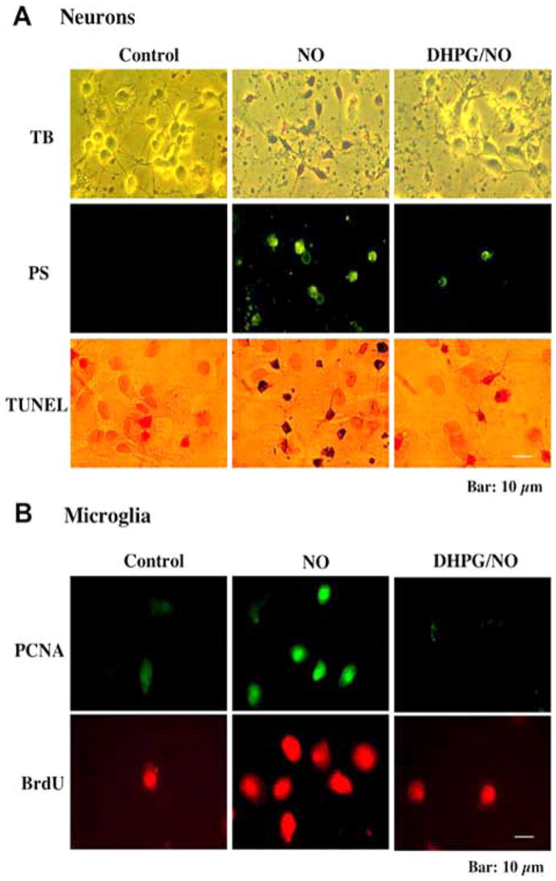
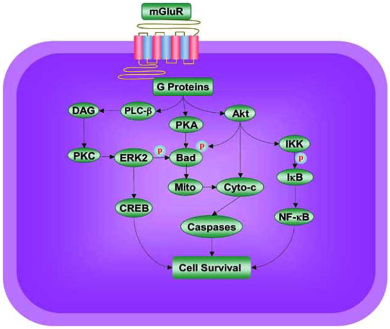
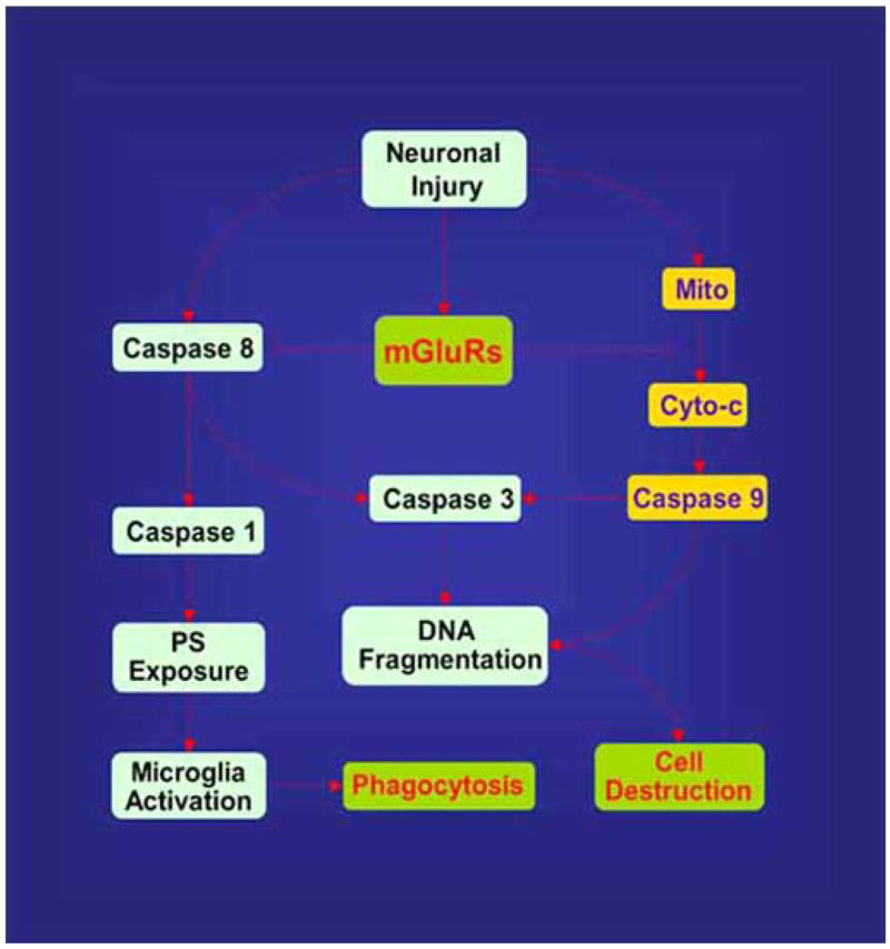
Metabotropic glutamate receptors (mGluRs) share a common molecular morphology with other G protein-linked receptors, but there expression throughout the mammalian nervous system places these receptors as essential mediators not only for the initial development of an organism, but also for the vital determination of a cell's fate during many disorders in the nervous system that include amyotrophic lateral sclerosis, Parkinson's disease, Alzheimer's disease, Huntington's disease, Multiple Sclerosis, epilepsy, trauma, and stroke. Given the ubiquitous distribution of these receptors, the mGluR system impacts upon neuronal, vascular, and glial cell function and is activated by a wide variety of stimuli that includes neurotransmitters, peptides, hormones, growth factors, ions, lipids, and light. Employing signal transduction pathways that can modulate both excitatory and inhibitory responses, the mGluR system drives a spectrum of cellular pathways that involve protein kinases, endonucleases, cellular acidity, energy metabolism, mitochondrial membrane potential, caspases, and specific mitogen-activated protein kinases. Ultimately these pathways can converge to regulate genomic DNA degradation, membrane phosphatidylserine (PS) residue exposure, and inflammatory microglial activation. As we continue to push the envelope for our understanding of this complex and critical family of metabotropic receptors, we should be able to reap enormous benefits for both clinical disease as well as our understanding of basic biology in the nervous system.


Figures

Fig. (1)
Fig. (1)
Activation of group I mGluRs by 3,5-dihydroxyphenylglycine (DHPG) maintains genomic DNA integrity and membrane phosphatidylserine (PS) asymmetry and prevents microglial activation. (A) Representative images illustrate cell membrane disruption with trypan blue staining, DNA fragmentation with terminal deoxynucleotidyl transferase nick end labeling (TUNEL), and phosphatidylserine (PS) exposure with annexin V phycoerythrin labeling in hippocampal neurons 24 hours following nitric oxide (NO) exposure (NOC-9, 300 μM). Pretreatment with the group I agonist DHPG (750 μM) 1 hour prior to the NO insult results in a significant reduction in trypan blue staining, DNA fragmentation, and membrane PS exposure. (B) Representative images of microglia are shown that illustrate increased micoglial activity assessed by proliferating cell nuclear antigen (PCNA) expression or the uptake of bromodeoxyuridine (BrdU) following the application of neuronal media that was exposed to either NO (NOC-9, 300 μM) or NO (NOC-9, 300 μM) plus DHPG. Administration of DHPG (750 μg/ml) one hour prior to neurons exposed to NO subsequently prevented PCNA expression and BrdU uptake in microglia illustrating that mGluR1 activation can block inflammatory microglial activation.
Fig. (2)
Fig. (2)
Potential signal transduction pathways of metabotropic glutamate receptors (mGluRs) that may foster cellular protection. mGluRs employ G-protein βγ to activate phospholipase β (PLC-β), diacylglycerol (DAG), and phosphoinositide 3 kinase (PI 3-K). These pathways lead to the activation of protein kinases A (PKA), B (Akt), and C (PKC). PKA has been shown to phosphorylate (p) Bad, a member of Bcl-2 protein family, which can prevent the induction of cell injury. Akt provides an anti-apoptotic survival signal through the phosphorylation and inactivation of Bad and stimulation of NF-kappaB (NF-κB) activity. Akt can activate IκB kinase (IKK) that can precipitate the phosphorylation (p) and degradation of IκB. This is followed by liberation of free NF-κB to promote cell survival. In regards to PKC, mGluRs can activate ERK2 through PKC. ERKs also may employ phosphorylation (p) of the pro-apoptotic protein Bad and induction of pro-survival gene expression via the cAMP responsive element-binding (CREB) protein dependent pathway to lead to cellular protection. mGluRs also preserve mitochondrial membrane potential (Mito) to block cytochrome c (Cyto-c) release and caspase activation that ultimately will lead to cellular demise.
Fig. (3)
Fig. (3)
Metabotropic glutamate receptors (mGluRs) prevent apoptotic neuronal injury, caspase induction, and microglial activation. Activation of mGluRs maintains mitochondrial membrane potential (Mito) to prevent the release of cytochrome c (Cyto c) and subsequent caspase activation. With the blockade of caspase activity, apoptotic DNA fragmentation and membrane phosphatidylserine (PS) exposure is prevented. The ability of mGluRs to block microglia activation relies upon the prevention of membrane phosphatidylserine (PS) exposure.

Similar articles

Cited by

References

    1. Aarts MM, Tymianski M. Novel treatment of excitotoxicity: targeted disruption of intracellular signalling from glutamate receptors. Biochem Pharmacol. 2003;66(6):877–86. - PubMed
    1. Aarts MM, Tymianski M. Molecular mechanisms underlying specificity of excitotoxic signaling in neurons. Curr Mol Med. 2004;4(2):137–47. - PubMed
    1. Abraham KE, McGinty JF, Brewer KL. The role of kainic acid/AMPA and metabotropic glutamate receptors in the regulation of opioid mRNA expression and the onset of pain-related behavior following excitotoxic spinal cord injury. Neuroscience. 2001;104(3):863–74. - PubMed
    1. Agrawal SK, Theriault E, Fehlings MG. Role of group I metabotropic glutamate receptors in traumatic spinal cord white matter injury. J Neurotrauma. 1998;15(11):929–41. - PubMed
    1. Albasanz JL, Dalfo E, Ferrer I, Martin M. Impaired metabotropic glutamate receptor/phospholipase C signaling pathway in the cerebral cortex in Alzheimer’s disease and dementia with Lewy bodies correlates with stage of Alzheimer’s-disease-related changes. Neurobiol Dis 2005 - PubMed

Publication types

MeSH terms

Substances