

Long-term follow-up of fixation patterns in eyes with central scotomas from geographic atrophy that is associated with age-related macular degeneration
- PMID: 16376656
- PMCID: PMC2528862
- DOI: 10.1016/j.ajo.2005.07.040
Long-term follow-up of fixation patterns in eyes with central scotomas from geographic atrophy that is associated with age-related macular degeneration
Abstract
Purpose: To study whether fixation patterns changed over time in patients with central scotomas from geographic atrophy from age-related macular degeneration.
Design: Prospective cohort study.
Methods: setting: Institutional. patient or study population: Prospective natural history study of geographic atrophy included 34 eyes of 25 patients with baseline acuity between 20/80 and 20/200 and with subsequent follow-up. observation procedures: Baseline and annual follow-up visits included best-corrected visual acuity, scanning laser ophthalmoscope macular perimetry, reading rate, and clinical evaluation. main outcome measures: Location of eccentric preferred retinal locus for fixation (PRL).
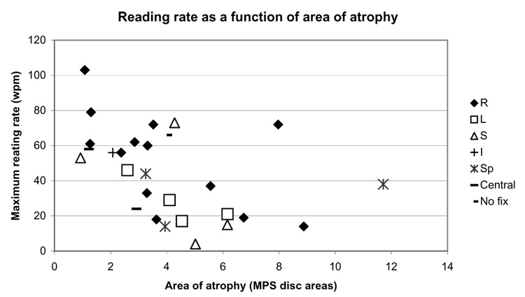
Results: At baseline, 77% of study eyes had a PRL. At the final visit (median follow-up, 5.3 years), 91% of study eyes had a PRL, with 81% of the eyes retaining the baseline PRL location. Fixation with the scotoma to the right and fixation with the scotoma superior were the first and second most common fixation patterns, respectively. Reading rates of <50 words/min were present in 54% of eyes. Eyes fixating with the scotoma to the left tended to have lower reading rates than eyes fixating with right or superior patterns.
Conclusion: Fixation with right pattern remained the most common fixation pattern, both in patients with a PRL at baseline and in patients who had a PRL during follow-up. Eyes with a PRL at baseline generally retained this pattern. The reading rate data suggest an advantage of fixation with right or superior pattern, rather than left. Reading rate declined further during follow-up in most patients.
Figures
Similar articles
-
Fixation patterns and reading rates in eyes with central scotomas from advanced atrophic age-related macular degeneration and Stargardt disease.Ophthalmology. 1996 Sep;103(9):1458-66. doi: 10.1016/s0161-6420(96)30483-1. Ophthalmology. 1996. PMID: 8841306 Free PMC article.
-
Improvement of visual acuity over time in patients with bilateral geographic atrophy from age-related macular degeneration.Retina. 2000;20(2):162-9. Retina. 2000. PMID: 10783949 Clinical Trial.
-
Preferred retinal locus development in patients with macular disease.Ophthalmology. 2005 Sep;112(9):1579-85. doi: 10.1016/j.ophtha.2005.03.027. Ophthalmology. 2005. PMID: 16087239
-
Evaluation of fixation pattern and reading ability in patients with Leber hereditary optic neuropathy.J Neuroophthalmol. 2013 Dec;33(4):344-8. doi: 10.1097/WNO.0b013e31829d1f5b. J Neuroophthalmol. 2013. PMID: 24256876
-
Preferred retinal loci and macular scotoma characteristics in patients with age-related macular degeneration.Can J Ophthalmol. 2005 Jun;40(3):303-12. doi: 10.1016/S0008-4182(05)80073-0. Can J Ophthalmol. 2005. PMID: 15947800 Review.
Cited by
-
The long-term natural history of geographic atrophy from age-related macular degeneration: enlargement of atrophy and implications for interventional clinical trials.Ophthalmology. 2007 Feb;114(2):271-7. doi: 10.1016/j.ophtha.2006.09.016. Ophthalmology. 2007. PMID: 17270676 Free PMC article.
-
Fixation Stability and Preferred Retinal Locus in Advanced Age-Related Macular Degeneration.Turk J Ophthalmol. 2022 Feb 23;52(1):23-29. doi: 10.4274/tjo.galenos.2021.27985. Turk J Ophthalmol. 2022. PMID: 35196836 Free PMC article.
-
Concise Review: Update on Retinal Pigment Epithelium Transplantation for Age-Related Macular Degeneration.Stem Cells Transl Med. 2019 May;8(5):466-477. doi: 10.1002/sctm.18-0282. Epub 2019 Feb 12. Stem Cells Transl Med. 2019. PMID: 30748126 Free PMC article. Review.
-
Development and Validation of Performance-Based Assessment of Daily Living Tasks in Age-Related Macular Degeneration.Transl Vis Sci Technol. 2024 Jun 3;13(6):9. doi: 10.1167/tvst.13.6.9. Transl Vis Sci Technol. 2024. PMID: 38884546 Free PMC article.
-
Geographic atrophy: Understanding the relationship between structure and function.Asia Pac J Ophthalmol (Phila). 2025 May-Jun;14(3):100207. doi: 10.1016/j.apjo.2025.100207. Epub 2025 May 19. Asia Pac J Ophthalmol (Phila). 2025. PMID: 40398512 Review.
References
-
- White JM, Bedell HE. The oculomotor reference in human with bilateral macular disease. Invest Ophthalmol Vis Sci. 1990;31:1149–1161. - PubMed
-
- Guez J-E, Gargasson J-FL, Rigaudiere F, O’Regan JK. Is there a systematic location for the pseudo-fovea in patients with central scotoma? Vision Res. 1993;9:1271–1279. - PubMed
-
- Acosta F, Lashkar K, Reynaud X, et al. Characterization of functional changes in macular holes and cysts. Ophthalmology. 1991;98:1820–1823. - PubMed
-
- Rohrschneider K, Gluck R, Blankenagel A, Volcker HE. Fixationsverhalten bei Morbus Stargardt. Ophthalmologe. 1997;94:624–628. - PubMed
Publication types
MeSH terms
Grants and funding
LinkOut - more resources
Full Text Sources
Medical
