. 2005 Dec 23;33(22):7000-10.
doi: 10.1093/nar/gki997. Print 2005.

Oligomerization of HEXIM1 via 7SK snRNA and coiled-coil region directs the inhibition of P-TEFb

Affiliations

Oligomerization of HEXIM1 via 7SK snRNA and coiled-coil region directs the inhibition of P-TEFb

Dalibor Blazek et al. Nucleic Acids Res. .

Abstract

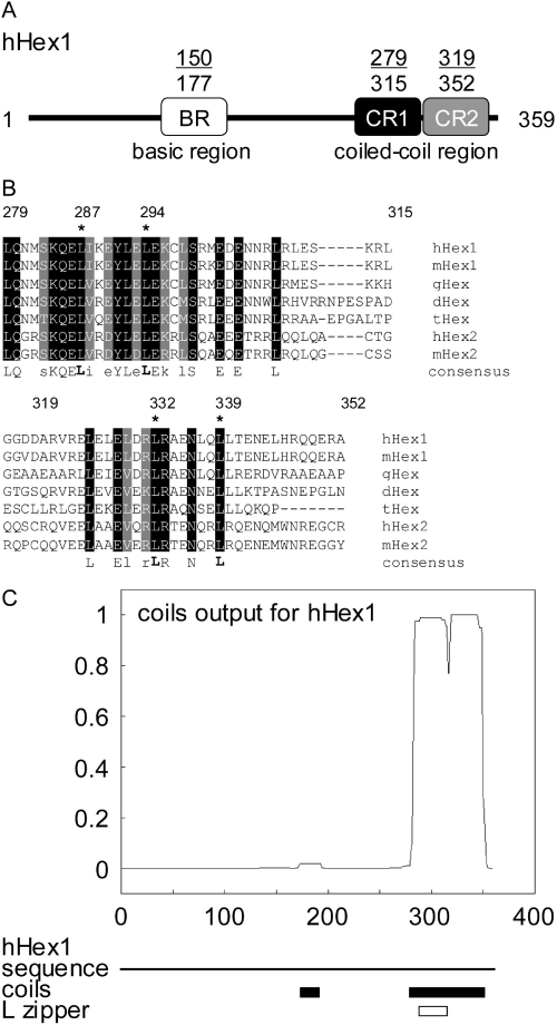
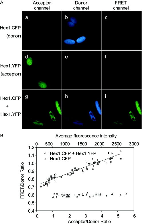
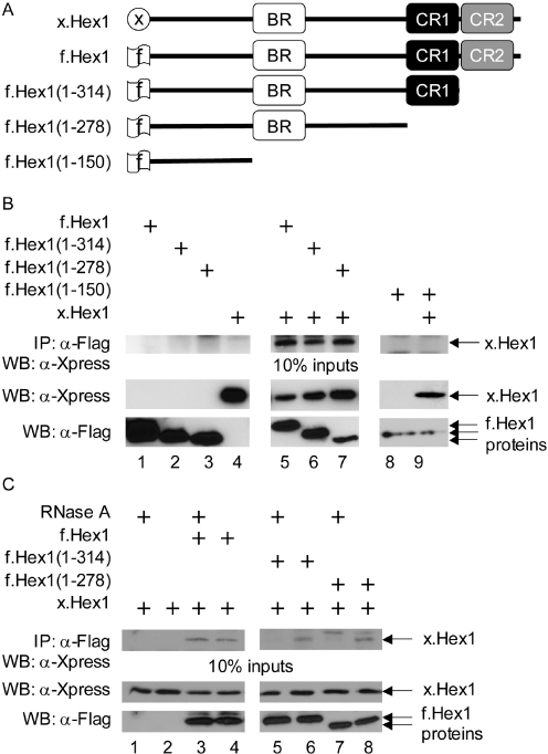
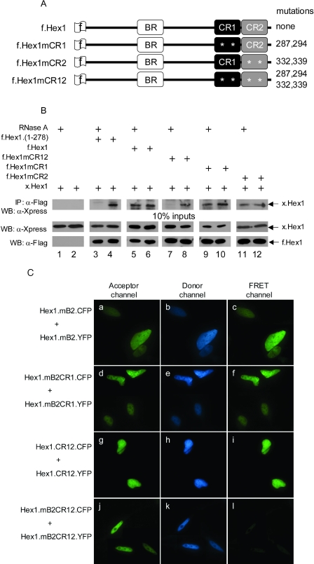
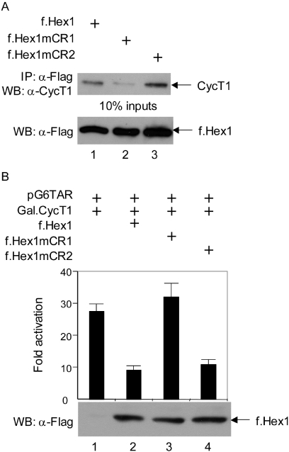
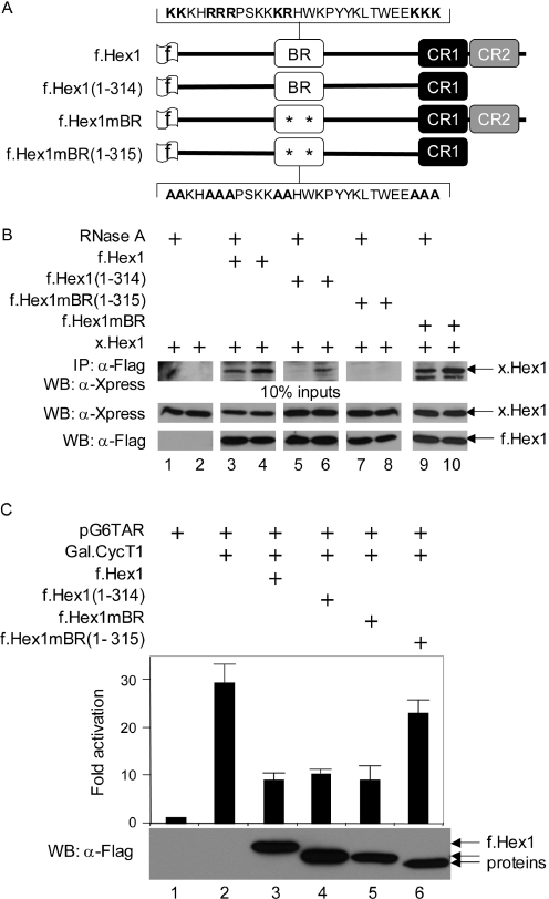
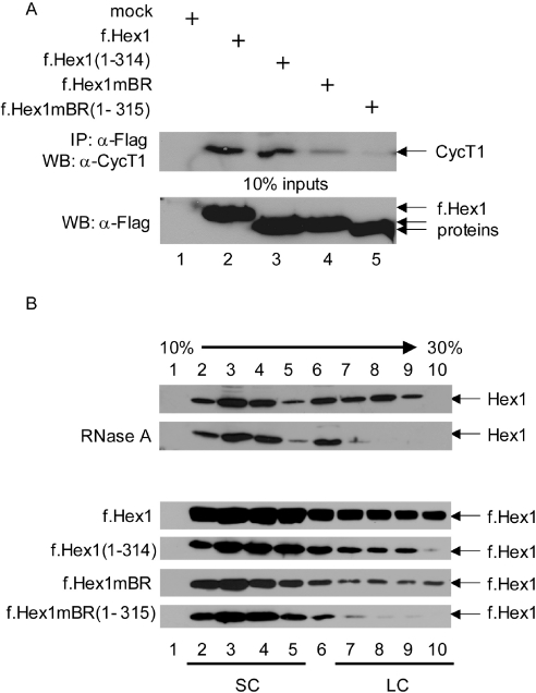
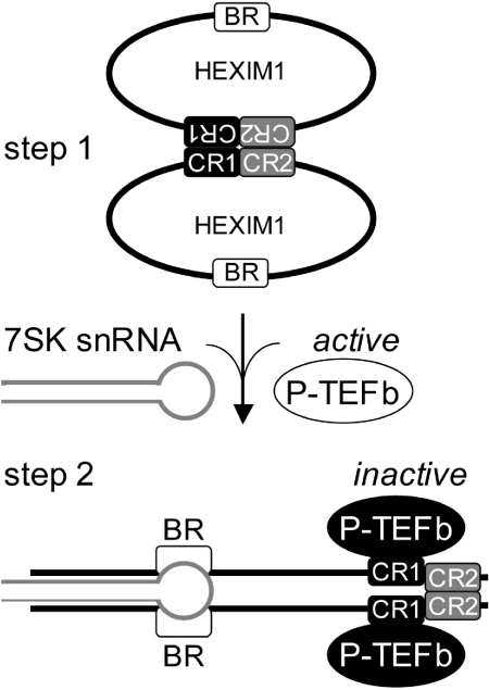
Transcriptional elongation of most eukaryotic genes by RNA polymerase II requires the kinase activity of the positive transcription elongation factor b (P-TEFb). The catalytically active P-TEFb complex becomes inactive when sequestered into the large complex by the cooperative actions of 7SK snRNA and HEXIM1. In this study, we report that HEXIM1 forms oligomers in cells. This oligomerization is mediated by its predicted coiled-coil region in the C-terminal domain and 7SK snRNA that binds a basic region within the central part of HEXIM1. Alanine-mutagenesis of evolutionary conserved leucines in the coiled-coil region and the digestion of 7SK snRNA by RNase A treatment prevent this oligomerization. Importantly, mutations of the N-terminal part of the coiled-coil region abrogate the ability of HEXIM1 to bind and inhibit P-TEFb. Finally, the formation of HEXIM1 oligomers via the C-terminal part of the coiled-coil or basic regions is critical for the inhibition of transcription. Our results suggest that two independent regions in HEXIM1 form oligomers to incorporate P-TEFb into the large complex and determine the inhibition of transcriptional elongation.


Figures

Figure 1
Figure 1
Prediction of CR1 and CR2 in the C-terminal domain of HEXIM1. (A) Schematic depiction of human HEXIM1 protein (hHex1). White box represents the BR. CR1 and CR2 are represented by black and gray boxes, respectively. The numbering above the boxes defines the boundaries of the regions. (B) Alignment of human (hHex1), mouse (mHex1), chicken (gHex1), zebrafish (dHex1), fish (tHex1) HEXIM1 and human (hHex2) and mouse (mHex2) HEXIM2 proteins. Black boxes indicate amino acid identity while shaded boxes the amino acid similarity. The numbering above the alignment corresponds to the boundaries of the predicted CR1 and CR2. Also, the numbered leucines, which are marked by an asterisk, were mutated to alanines. The consensus sequence is indicated below the alignment. (C) The graph indicates the probability of coiled-coil for the human HEXIM1 protein as predicted by the coils prediction program. The line below the graph corresponds to the sequence of hHex1 and the boxes indicate regions with high probability of coiled-coil (black box) and leucine zipper (white box) as predicted by the coils software.
Figure 2
Figure 2
HEXIM1 oligomerizes in the nucleus of cells. (A) Representative images of Hex1.YFP and Hex1.CFP chimeras, which were expressed alone or together in cells. Amounts of nuclear fluorescence were quantified in the yellow, cyan and FRET channels. Energy transfer resulted in an increased FRET at the expense of donor emissions (FRET/donor) in the co-expressing cells relative to the cells containing the donors alone. (B) FRET/donor ratio increased proportionally with the amount of the acceptor relative to the donor in cells co-expressing Hex1.YFP and Hex1.CFP chimeras. The slope of the graph represents the extent of FRET at normalized acceptor/donor levels. There was no FRET between Hex1.CFP proteins expressed alone.
Figure 3
Figure 3
The C-terminal domain and 7SK snRNA mediate the oligomerization of HEXIM1. (A) Schematic diagram of Hex1 proteins used. The signs at their N-termini depict the respective tags. (B) HEXIM1 forms oligomers. The x.Hex1 and f.Hex1 proteins were either expressed alone (lanes 4 and 1, 2, 3, 8, respectively) or f.Hex1 was co-expressed with x.Hex1 in HeLa cells (lanes 5–7 and 9) as indicated. Lysates were co-immunoprecipitated with anti-FLAG agarose beads and immunoprecipitates of x.Hex1 were identified as presented on the upper western blot (WB). The middle and lower WB contain 10% of input proteins for immunoprecipitations (IP). Wild-type and mutant HEXIM1 proteins are identified by arrows. (C) 7SK snRNA and the C-terminal domain of HEXIM1 mediate the oligomerization of HEXIM1. x.Hex1 was expressed alone (lanes 1 and 2) or with the indicated f.Hex1 proteins (lanes 3–8). IP were performed as in (B) and were treated with RNase A where indicated.
Figure 4
Figure 4
Combined disruptions of the CR and BR abolish HEXIM1 oligomerization. (A) Schematic diagram of Hex1 proteins used. The sign at their N-termini depicts the FLAG tag. The asterisks within the CR1 and CR2 indicate the mutations of leucines to alanines. The numbering indicates the positions of the mutated leucines in f.Hex1 proteins. (B) HEXIM1 with the disrupted CR does not oligomerize in the absence of 7SK snRNA. Proteins with the disrupted CR1 or CR2 (lanes 9–12) or CR (lanes 7 and 8) were co-expressed with x.Hex1 in HeLa cells. The lysates were treated with RNase A where indicated, immunoprecipitated with anti-FLAG agarose beads and immunoprecipitates were subjected to SDS–PAGE and WB (upper panel). Lower panels represent 10% input of proteins. (C) Combined disruptions of the CR and the 7SK snRNA binding site abolish completely HEXIM1 oligomerization in cells. FRET analysis was performed as in Figure 2. Representative images of the nuclei in which Hex1.YFP and Hex1.CFP mutant proteins were co-expressed are presented. The amounts of nuclear fluorescence were quantified in the yellow, cyan and FRET channels.
Figure 5
Figure 5
CR1 in HEXIM1 is required for its binding to P-TEFb and inhibition of transcription. (A) Disruption of the CR1 abrogates P-TEFb binding. f.Hex1 proteins were expressed in HeLa cells (6 µg, lanes 1–3) and immunoprecipitated with anti-FLAG agarose beads. Amounts of bound endogenous CycT1 is presented by WB (upper panel). The amounts of f.Hex1 proteins are indicated in WB below (lower panel). (B) f.Hex1mCR1 does not inhibit transcription. HeLa cells expressed pG6TAR (0.3 µg), Gal.CycT1 (1 µg) and f.Hex1 (2.7 µg) as depicted. Bars correspond to CAT values and the lower panel presents expression (WB) of f.Hex1 as indicated by the arrow.
Figure 6
Figure 6
Oligomerization of HEXIM1 via its BR or CR2 is required for the inhibition of transcription. (A) Schematic diagram of Hex1 proteins used. The BR, CR1 and CR2 regions participating in the oligomerization are depicted. The wild-type and the mutated residues of the BR are depicted above and below the diagram, respectively. The mutated BR is indicated by asterisk. The schematic picture represents the f.Hex1 and mutant f.Hex1(1–314), f.Hex1mBR and f.Hex1mBR(1–315) proteins used. (B) HEXIM1 without the BR and the CR2 does not oligomerize. The x.Hex1 and f.Hex1 proteins were co-expressed as depicted. Lysates were treated with RNase A where noted and IP was performed as described. Upper panel represents WB with the immunoprecipitated x.Hex1 proteins, whereas the middle and lower panels show 10% input of proteins used for IP. (C) HEXIM1 without the BR and the CR2 does not inhibit P-TEFb. Bars represent CAT data obtained by co-transfection of HeLa cells with pG6TAR (0.3 µg), Gal.CycT1 (1 µg) and indicated f.Hex1 plasmids (2.7 µg). The lower panel presents the expression of f.Hex1 proteins.
Figure 7
Figure 7
Oligomerization of HEXIM1 via BR or CR2 is required for the incorporation of HEXIM1 into the LC of P-TEFb. (A) HEXIM1 with disrupted BR and CR2 does not bind P-TEFb. HeLa cells were either mock transfected (lane 1) or transfected with f.Hex1 plasmids (6 µg, lanes 2–5). The f.Hex1 proteins were immunoprecipitated with anti-FLAG agarose beads. Amounts of bound endogenous CycT1 is presented by WB (upper panel). The amounts of f.Hex1 proteins are indicated in WB below (lower panel). (B) HEXIM1 with disrupted BR and CR2 does not incorporate into the LC. Lysates from either mock or f.Hex1 plasmids transfected cells were divided into ten fractions by glycerol gradient centrifugation. Amounts of endogenous HEXIM1 proteins in each fraction were analyzed by immunoblotting as shown on the panels. The numbers below and above panels depict the glycerol fractions and arrow indicates increasing glycerol gradient (from 10 to 30%). SC and LC stand for small complex and large complex of the P-TEFb, respectively.
Figure 8
Figure 8
A model how the oligomerization of HEXIM1 via 7SK snRNA and CR directs the incorporation of P-TEFb into the LC. In the absence of 7SK snRNA, free HEXIM1 proteins are oligomers (two ovals, step 1). This oligomerization is mediated solely by the CR1 and CR2 (black and gray rectangles, respectively) in the C-terminal domain of HEXIM1. The BR is depicted as a white rectangle. The small complex of P-TEFb (white oval) is active. In contrast, when 7SK snRNA (hairpin) binds the BRs in HEXIM1, it facilitates the second oligomerization event. Subsequently, P-TEFb is incorporated into the LC, leading to its inactivation (black ovals, step 2). In this complex, the CR1 in HEXIM1 mediates the binding to P-TEFb, whereas the CR2 facilitates HEXIM1 oligomerization.

References

    1. Sims R.J., III, Belotserkovskaya R., Reinberg D. Elongation by RNA polymerase II: the short and long of it. Genes Dev. 2004;18:2437–2468. - PubMed
    1. Barboric M., Peterlin B.M. A new paradigm in eukaryotic biology: HIV Tat and the control of transcriptional elongation. PLoS Biol. 2005;3:e76. - PMC - PubMed
    1. Price D.H. P-TEFb, a cyclin-dependent kinase controlling elongation by RNA polymerase II. Mol. Cell. Biol. 2000;20:2629–2634. - PMC - PubMed
    1. Shim E.Y., Walker A.K., Shi Y., Blackwell T.K. CDK-9/cyclin T (P-TEFb) is required in two postinitiation pathways for transcription in the C.elegans embryo. Genes Dev. 2002;16:2135–2146. - PMC - PubMed
    1. Chao S.H., Price D.H. Flavopiridol inactivates P-TEFb and blocks most RNA polymerase II transcription in vivo. J. Biol. Chem. 2001;276:31793–31799. - PubMed

Publication types

Substances