. 2006 Jan;80(2):697-709.
doi: 10.1128/JVI.80.2.697-709.2006.

Kaposi's sarcoma-associated herpesvirus-encoded latency-associated nuclear antigen induces chromosomal instability through inhibition of p53 function

Affiliations

Kaposi's sarcoma-associated herpesvirus-encoded latency-associated nuclear antigen induces chromosomal instability through inhibition of p53 function

Huaxin Si et al. J Virol. 2006 Jan.

Abstract

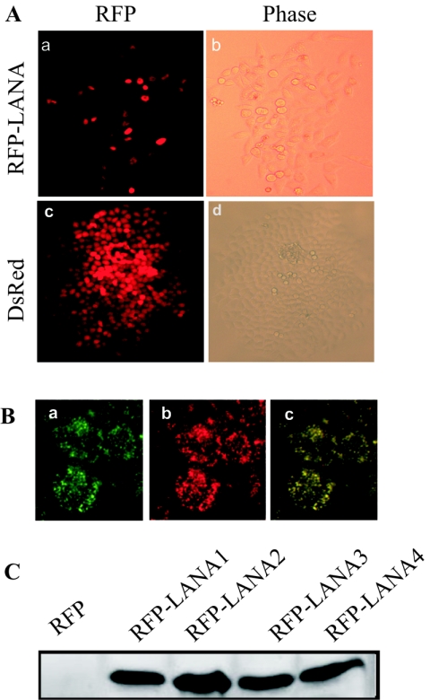
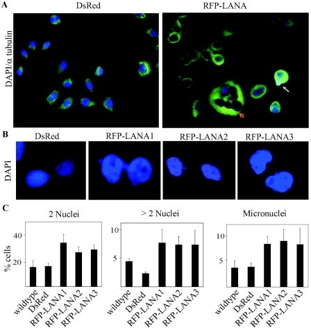
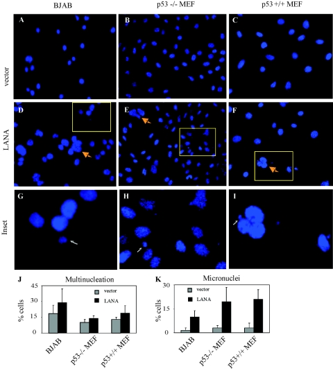
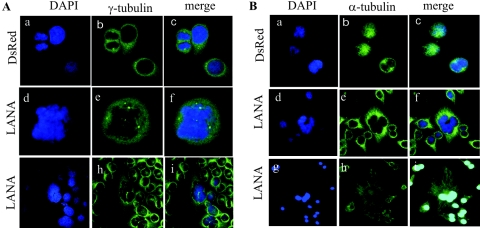
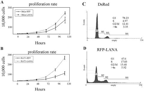
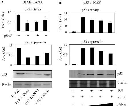
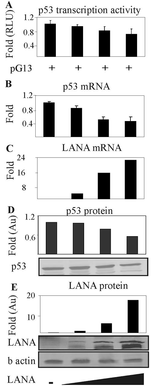
Kaposi's sarcoma-associated herpesvirus (KSHV) is predominantly associated with three human malignancies, KS, primary effusion lymphoma, and multicentric Castleman's disease. These disorders are linked to genomic instability, known to be a crucial component of the oncogenic process. Latency-associated nuclear antigen (LANA), encoded by open reading frame 73 of the KSHV genome, is a latent protein consistently expressed in all KSHV-associated diseases. LANA is important in viral genome maintenance and is associated with cellular and viral proteins to regulate viral and cellular gene expression. LANA interacts with the tumor suppressor genes p53 and pRb, indicating that LANA may target these proteins and promote oncogenesis. In this study, we generated cell lines which stably expressed LANA to observe the effects of LANA expression on cell phenotype. LANA expression in these stable cell lines showed a dramatic increase in chromosomal instability, indicated by the presence of increased multinucleation, micronuclei, and aberrant centrosomes. In addition, these stable cell lines demonstrated an increased proliferation rate and as well as increased entry into S phase in both stable and transiently transfected LANA-expressing cells. Additionally, p53 transcription and its transactivation activity were suppressed by LANA expression in a dose-dependent manner. LANA may therefore promote chromosomal instability by suppressing the functional activities of p53, thereby facilitating KSHV-mediated pathogenesis and cancer.


Figures

FIG. 1.
FIG. 1.
Generation of cells with stable expression of KSHV LANA. (A) Nuclear expression of RFP-LANA (a) and in a HeLa colony (b) and whole-cell expression of pDsRed1 (c) and in a HeLa colony (d). (B) Immunocolocalization of LANA and RFP in stable cells. Cells expressing LANA were fixed in 1:1 methanol-acetone and incubated with an anti-LANA serum. (a) LANA was localized by use of an Alexa Fluor-conjugated secondary antibody. (b) RFP was directly visualized under a fluorescence microscope. (c) The merged image shows the colocalization of those proteins in the nuclear. (C) LANA expression in stable cells was monitored by Western blot analysis with an anti-LANA serum. LANA expression was detected in RFP-LANA colonies but not in the RFP colony.
FIG. 2.
FIG. 2.
Induction of multinucleation, micronuclei, and chromosomal laggings in LANA-expressing HeLa cells. (A) DAPI staining in representative fields shows increased multinucleation (yellow arrows) and numbers of micronuclei (white arrows) in cells stably expressing LANA in comparison with cells stably expressing the RFP vector. Cells were also counterstained with α-tubulin to show the morphological changes. (B) Presence of a mitotic bridge in cells stably expressing LANA but not in cells stably expressing the RFP vector. (C) Quantification of multinucleation (two nuclei and more than two nuclei) and micronuclei in HeLa cells as well as HeLa cells with the RFP vector and RFP-LANA expression. DsRed, pDsRed1.
FIG. 3.
FIG. 3.
Induction of multinucleation and micronuclei in LANA-expressing Rat1 cells. (A) DAPI staining of representative fields shows increased multinucleation (yellow arrows) and numbers of micronuclei (white arrows) in cells stably expressing LANA. (B) Quantification of Rat1 cells with two nuclei in each cell line. (C) Quantification of Rat1 cells with more than two nuclei in each cell line. (D) Quantification of Rat1 cells with micronuclei in each cell line.
FIG. 4.
FIG. 4.
Induction of multinucleation and micronuclei in LANA-expressing cells. BJAB cells, p53−/− MEF, and p53+/+ MEF were transfected with a LANA construct and subjected to G418 selection. (A-C) DAPI staining of BJAB cells (A), p53−/− MEF (B), and p53+/+ MEF (C) with mock control DNA. (D-F) DAPI staining of representative fields shows increased multinucleation (yellow arrows) and numbers of micronuclei (white arrows) in BJAB cells (D), p53−/− MEF (E), and p53+/+ MEF (F) with LANA stably expressed. (G-I) Inset fields show micronuclei (white arrows) corresponding to the yellow rectangles in panels D to F. (J-K) Quantification of cells with more than two nuclei (J) and micronuclei (K).
FIG. 5.
FIG. 5.
Induction of multinucleation and micronuclei in Rat1 cells and MEF with LANA and LANA derivatives. Rat1 cells and p53−/− and p53+/+ MEF were transfected with full-length LANA and LANA-derivative constructs and subjected to selection for 3 weeks. DAPI staining of representative fields shows multinucleation (yellow arrows) in Rat1 cells with the mock control, N-terminal LANA expression, C-terminal LANA expression, and full-length LANA expression, and inset fields show micronuclei (white arrows) corresponding to the yellow rectangles in the upper panel (A). (B-C) Quantification of cells with more than two nuclei (B) and micronuclei (C). (D-E) Staining of representative fields shows nuclear morphological changes in p53−/− MEF (D) and p53+/+ MEF (E) with a mock control, N-terminal LANA expression, C-terminal LANA expression, and full-length LANA expression. (F-G) Quantification of cells with at least two nuclei and micronuclei in p53−/− MEF (F) and p53+/+ MEF (G).
FIG. 6.
FIG. 6.
Induction of abnormal mitotic spindle poles and centrosomes in LANA-expressing cells. (A) DAPI (a, d, and g) and γ-tubulin (b, e and h) staining in representative fields shows increased centrosome numbers in LANA expression cells (d-i) but not in RFP expression cells (a-c). (B) DAPI (a, d, and g) and α-tubulin (b, e and h) staining in representative fields shows abnormal spindle poles in LANA-expressing cells (d-i) but not in RFP expression cells (a-c).
FIG. 7.
FIG. 7.
High proliferation rate and replication rate of LANA-expressing cells in comparison to that of mock control cells. A total of 3 × 10⁴ HeLa and 4 × 10⁴ stable Rat1 cells were seeded on day zero, and absolute cell counts were determined daily for the next 5 days. (A) Proliferative rate of stable HeLa cells. (B) Proliferate rate of stable Rat1 cells. Values shown represent the means plus standard deviations from three separate experiments. To determine possible changes in cell cycle profile due to LANA, the cellular DNA content was measured by propidium iodide staining and flow cytometry. (C) Cell cycle profile of vector control Rat1 cells. (D) Cell cycle profile of LANA-expressing Rat1 cells.
FIG. 8.
FIG. 8.
p53 transcription activity is repressed in cells stably expressing LANA and transiently expressing cells. To test the transcription activity of p53, 1 μg of the pG13 plasmid, which contains tandem repeats of the p53-binding site, was transfected into stable BJAB-RFP and BJAB-LANA cells. In p53−/− cells, 10 μg of the p53 construct was introduced in addition to LANA and reporter constructs. Cells were harvested 15 h posttransfection, and luciferase expression was measured as described in Materials and Methods. p53 transcription activity and the expression of p53 were tested by the lysate of 2 million transfected cells with anti-p53 and anti-β actin antibodies in cells stably expressing LANA (A) and in MEF transiently expressing LANA. Au, arbitrary units; Rlu, relative light units.
FIG. 9.
FIG. 9.
p53 transcription activity is repressed in cells transiently expressing LANA in a dose-dependent manner. To test the effect of LANA expression on the transcription activity of p53, increased amounts of the pA3M-LANA (0, 2, 5, 10 μg) plasmid as well as 1 μg of the pG13 plasmid was introduced into BJAB cells. Cells were harvested 18 h posttransfection, and luciferase expression was measured as described in Materials and Methods. (A) mRNA expression of p53 and LANA was determined by real-time RT-PCR (B and C), and the protein expression of p53 and LANA was determined by Western blotting (D and E). Au, arbitrary units; RLU, relative light units.
FIG. 10.
FIG. 10.
Model of the mechanism for LANA to facilitate chromosomal instability. Upon KSHV latent infection, the activation of viral genes (such as LANA, v-cyclin, and v-FLIP) accompanied by spontaneous errors and inheritance led to an aberrant replication structure and DNA damage. These aberrations can trigger the activation of p53 and concurrent growth arrest and apoptosis. However, suppression of p53 transcripts levels by LANA expression leads to a failure to monitor the generated aberrations by p53-dependent checkpoints, thus resulting in the formation of a polyploid/aneuploid population of cells with amplified numbers of centrosomes. This may allow the accumulation of chromosomal instability and additional genetic changes that ultimately result in a fully malignant phenotype.

References

    1. An, F. Q., N. Compitello, E. Horwitz, M. Sramkoski, E. S. Knudsen, and R. Renne. 2005. The latency-associated nuclear antigen of Kaposi's sarcoma-associated herpesvirus modulates cellular gene expression and protects lymphoid cells from p16INK4A-induced cell cycle arrest. J. Biol. Chem. 280:3862-3874. - PubMed
    1. Attardi, L. D. 2005. The role of p53-mediated apoptosis as a crucial anti-tumor response to genomic instability: lessons from mouse models. Mutat. Res. Fund. Mol. Mech. Mutagen. 569:145-157. - PubMed
    1. Ballestas, M. E., P. A. Chatis, and K. M. Kaye. 1999. Efficient persistence of extrachromosomal KSHV DNA mediated by latency-associated nuclear antigen. Science 284:641-644. - PubMed
    1. Ballestas, M. E., and K. M. Kaye. 2001. Kaposi's sarcoma-associated herpesvirus latency-associated nuclear antigen 1 mediates episome persistence through cis-acting terminal repeat (TR) sequence and specifically binds TR DNA. J. Virol. 75:3250-3258. - PMC - PubMed
    1. Blagosklonny, M. V., P. Giannakakou, L. Y. Romanova, K. M. Ryan, K. H. Vousden, and T. Fojo. 2001. Inhibition of HIF-1- and wild-type p53-stimulated transcription by codon Arg175 p53 mutants with selective loss of functions. Carcinogenesis 22:861-867. - PubMed

Publication types