

Epstein-Barr virus (EBV) genome and expression in breast cancer tissue: effect of EBV infection of breast cancer cells on resistance to paclitaxel (Taxol)
- PMID: 16378986
- PMCID: PMC1346837
- DOI: 10.1128/JVI.80.2.845-853.2006
Epstein-Barr virus (EBV) genome and expression in breast cancer tissue: effect of EBV infection of breast cancer cells on resistance to paclitaxel (Taxol)
Abstract
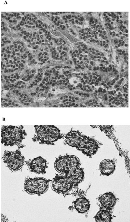
The Epstein-Barr virus (EBV) has been detected in subsets of breast cancers. In order to elaborate on these observations, we quantified by real-time PCR (Q-PCR) the EBV genome in biopsy specimens of breast cancer tissue as well as in tumor cells isolated by microdissection. Our findings show that EBV genomes can be detected by Q-PCR in about half of tumor specimens, usually in low copy numbers. However, we also found that the viral load is highly variable from tumor to tumor. Moreover, EBV genomes are heterogeneously distributed in morphologically identical tumor cells, with some clusters of isolated tumor cells containing relatively high genome numbers while other tumor cells isolated from the same specimen may be negative for EBV DNA. Using reverse transcription-PCR, we detected EBV gene transcripts: EBNA-1 in almost all of the EBV-positive tumors and RNA of the EBV oncoprotein LMP-1 in a smaller subset of the tissues analyzed. Moreover, BARF-1 RNA was detected in half of the cases studied. Furthermore, we observed that in vitro EBV infection of breast carcinoma cells confers resistance to paclitaxel (taxol) and provokes overexpression of a multidrug resistance gene (MDR1). Consequently, even if a small number of breast cancer cells are EBV infected, the impact of EBV infection on the efficiency of anticancer treatment might be of importance.
Figures


References
-
- Baker, E. K., R. W. Johnstone, J. R. Zalcberg, and A. El-Osta. 8. August 2005. Epigenetic changes to the MDR1 locus in response to chemotherapeutic drugs. Oncogene 10.1038/sj.onc.1208955. - PubMed
-
- Blankenstein, M. A. 1990. Comparison of ligand binding assay and enzyme immunoassay of oestrogen receptor in human breast cancer cytosols. Experience of the E.O.R.T.C. Receptor Group. Breast Cancer Res. Treat. 17:91-98. - PubMed
-
- Bonnet, M., J. M. Guinebretiere, E. Kremmer, V. Grunewald, E. Benhamou, G. Contesso, and I. Joab. 1999. Detection of Epstein-Barr virus in invasive breast cancers. J. Natl. Cancer Inst. 91:1376-1381. - PubMed
-
- Brengel-Pesce, K., P. Morand, A. Schmuck, M. J. Bourgeat, M. Buisson, G. Bargues, M. Bouzid, and J. M. Seigneurin. 2002. Routine use of real-time quantitative PCR for laboratory diagnosis of Epstein-Barr virus infections. J. Med. Virol. 66:360-369. - PubMed
Publication types
MeSH terms
Substances
LinkOut - more resources
Full Text Sources
Other Literature Sources
Medical
