. 2006 Jan;80(2):1015-24.
doi: 10.1128/JVI.80.2.1015-1024.2006.

Low pH-dependent endosomal processing of the incoming parvovirus minute virus of mice virion leads to externalization of the VP1 N-terminal sequence (N-VP1), N-VP2 cleavage, and uncoating of the full-length genome

Affiliations

Low pH-dependent endosomal processing of the incoming parvovirus minute virus of mice virion leads to externalization of the VP1 N-terminal sequence (N-VP1), N-VP2 cleavage, and uncoating of the full-length genome

Bernhard Mani et al. J Virol. 2006 Jan.

Abstract

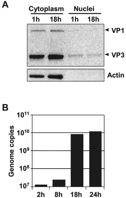
Minute virus of mice (MVM) enters the host cell via receptor-mediated endocytosis. Although endosomal processing is required, its role remains uncertain. In particular, the effect of low endosomal pH on capsid configuration and nuclear delivery of the viral genome is unclear. We have followed the progression and structural transitions of DNA full-virus capsids (FC) and empty capsids (EC) containing the VP1 and VP2 structural proteins and of VP2-only virus-like particles (VLP) during the endosomal trafficking. Three capsid rearrangements were detected in FC: externalization of the VP1 N-terminal sequence (N-VP1), cleavage of the exposed VP2 N-terminal sequence (N-VP2), and uncoating of the full-length genome. All three capsid modifications occurred simultaneously, starting as early as 30 min after internalization, and all of them were blocked by raising the endosomal pH. In particles lacking viral single-stranded DNA (EC and VLP), the N-VP2 was not exposed and thus it was not cleaved. However, the EC did externalize N-VP1 with kinetics similar to those of FC. The bulk of all the incoming particles (FC, EC, and VLP) accumulated in lysosomes without signs of lysosomal membrane destabilization. Inside lysosomes, capsid degradation was not detected, although the uncoated DNA of FC was slowly degraded. Interestingly, at any time postinfection, the amount of structural proteins of the incoming virions accumulating in the nuclear fraction was negligible. These results indicate that during the early endosomal trafficking, the MVM particles are structurally modified by low-pH-dependent mechanisms. Regardless of the structural transitions and protein composition, the majority of the entering viral particles and genomes end in lysosomes, limiting the efficiency of MVM nuclear translocation.


Figures

FIG. 1.
FIG. 1.
Kinetics of N-VP2 cleavage. (A) A9 cells (105) were infected with nonpurified MVM stocks (one PFU per cell) without detectable levels of VP3. At different intervals postinfection, the viral structural proteins were analyzed by 10% PAGE. After transfer to a polyvinylidene difluoride membrane, the blot was probed with a rabbit anti-VP antibody, followed by a horseradish peroxidase-conjugated secondary antibody. (B) Western blot analysis of purified FC and EC at 2 h postinfection. (C) A9 cells were infected with MVM stocks as indicated above. At different intervals postinfection, the cleavage of VP2 was examined by immunofluorescence with an antibody against capsids (red) and antibody against the N-VP2 (green).
FIG. 2.
FIG. 2.
Kinetics of N-VP1 externalization. (A) Externalization of N-VP1 from full-virus capsids was examined by immunofluorescence at various times postinfection. Antibody against capsids (green) and antibody against the N-VP1 (red) are shown. (B) Externalization of N-VP1 from empty capsids was examined at 2 h postinfection.
FIG. 3.
FIG. 3.
Detection of full-length genome externalization during the endosomal trafficking of MVM. (A) MVM genomic regions covered by the probes are shadowed. (B) A9 cells (105) seeded onto coverslips were infected with MVM as specified in Materials and Methods. At various times postinfection, the presence of externalized viral DNA was examined by in situ hybridization with probes covering the 3′ or 5′ regions of the MVM genome (red) and the viral capsids were detected with the MAb B7 against assembled virions (green). (C) Negative controls: cells infected with FC and hybridization with an unrelated MVM probe (derived from B19 sequences). Cells inoculated with empty particles and hybridization with the 5′ MVM probe. (D) Detection of exposed viral DNA at 21 h postinfection in quiescent cells.
FIG. 4.
FIG. 4.
Effect of chloroquine on the N-VP2 cleavage, N-VP1 externalization and uncoating. A9 cells (105) were infected with MVM. At 6 h postinfection, the presence of N-VP2, N-VP1, and the 5′ region of the viral genome was examined by immunofluorescence in the presence or absence of chloroquine (100 μM). The virus capsids were detected with the MAb B7 (against assembled capsids).
FIG. 5.
FIG. 5.
Blocking of VP2 cleavage in vivo. (A) Effect of endosome- and proteasome-disturbing agents on VP2 cleavage. A9 cells were infected with MVM stocks without VP3 in the presence of one of the following inhibitors: BA (150 nM), CHLO (100 μM), BFA (5 μg/ml), MG132 (25 μM), or aclarubicin (0.1 μM). (B) Effect of protease inhibitors on the VP2 cleavage. A9 cells were infected in the presence of one of the following inhibitors: TPCK (20 μM), TLCK (20 μM), aprotinin (2 μg/ml), E64 (10 μM), or pepstatin A (1 μM). At 0, 2, and 24 h postinternalization, cells were lysed in protein loading buffer and viral proteins were detected. NT, nontreated.
FIG. 6.
FIG. 6.
Effect of incoming full-virus, empty capsids, and virus-like particles on lysosomal membrane integrity. (A) The integrity of the lysosomal membranes from A9 cells infected with full-virus capsids, empty capsids, or VLP was examined with the 10-kDa fluid-phase lysosomal marker dextran Texas Red (Dex-10; red). The integrity of the viral capsids was examined with the MAb B7 against assembled capsids (green). (B) Externalization of N-VP1 and cleavage of N-VP2 examined at 18 h p.i. in cells infected with FC.
FIG. 7.
FIG. 7.
Cytoplasmic and nuclear distribution of radiolabeled viral proteins. (A) A9 cells were infected with radiolabeled full-virus capsids (1 PFU per cell). After 1 h (background control for viral nuclear accumulation) and 18 h p.i., nuclei and cytoplasm were separated and the viral proteins present in the nuclear and cytoplasmic fractions (nuclei/cytoplasm ratio, 1:1) were examined. (B) DNA replication kinetics of the radiolabeled full-virus capsids. A9 cells were infected (1 PFU per cell). After 2, 8, 18, and 24 h p.i., the viral DNA was quantified by real-time PCR.
FIG. 8.
FIG. 8.
Schematic representation of the capsid rearrangements during the endosomal trafficking of MVM virions (FC), EC, and VLP. The amounts and ratio of the represented viral proteins do not correspond to the amounts and ratio in the natural capsid. p, phosphorylated; np, nonphosphorylated. The symbols are the same as those used in reference 28.

Similar articles

Cited by

References

    1. Bantel-Schaal, U., B. Hub, and J. Kartenbeck. 2002. Endocytosis of adeno-associated virus type 5 leads to accumulation of virus particles in the Golgi compartment. J. Virol. 76:2340-2349. - PMC - PubMed
    1. Bartlett, J. S., R. Wilcher, and R. J. Samulski. 2000. Infectious entry pathway of adeno-associated virus and adeno-associated virus vectors. J. Virol. 74:2777-2785. - PMC - PubMed
    1. Basak, S., and H. Turner. 1992. Infectious entry pathway for canine parvovirus. Virology 186:368-376. - PubMed
    1. Bleker, S., F. Sonntag, and J. A. Kleinschmidt. 2005. Mutational analysis of narrow pores at the fivefold symmetry axes of adeno-associated virus type 2 capsids reveals a dual role in genome packaging and activation of phospholipase A2 activity. J. Virol. 79:2528-2540. - PMC - PubMed
    1. Canaan, S., Z. Zadori, F. Ghomashchi, J. Bollinger, M. Sadilek, M. E. Moreau, P. Tijssen, and M. H. Gelb. 2004. Interfacial enzymology of parvovirus phospholipases A2. J. Biol. Chem. 279:14502-14508. - PubMed

Publication types

LinkOut - more resources