

Does amygdalar perfusion correlate with antidepressant response to partial sleep deprivation in major depression?
- PMID: 16380239
- PMCID: PMC2468214
- DOI: 10.1016/j.pscychresns.2005.09.007
Does amygdalar perfusion correlate with antidepressant response to partial sleep deprivation in major depression?
Abstract
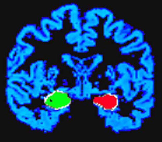
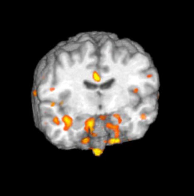
This study used functional MRI (fMRI) to clarify the sites of brain activity associated with the antidepressant effects of sleep deprivation (SD). We hypothesized: (1) baseline perfusion in right and left amygdalae will be greater in responders than in nonresponders; (2) following partial sleep deprivation (PSD), perfusion in responders' right and left amygdalae would decrease. Seventeen unmedicated outpatients with current major depression and eight controls received perfusion-weighted fMRI and structural MRI at baseline and following 1 night of late-night PSD. Baseline bilateral amygdalar perfusion was greater in responders than nonresponders. Clusters involving both amygdalae decreased from baseline to PSD specifically in responders. Right amygdalar perfusion diverged with PSD, increasing in nonresponders and decreasing in responders. These novel amygdalar findings are consistent with the overarousal hypothesis of SD as well as other functional imaging studies showing increased baseline amygdalar activity in depression and decreased amygdalar activity with remission or antidepressant medications.
Figures
References
-
- Abercrombie HC, Schaefer SM, Larson CL, Oakes TR, Lindgren KA, Holden JE, Perlman SB, Turski PA, Krahn DD, Benca RM, Davidson RJ. Metabolic rate in the right amygdala predicts negative affect in depressed patients. NeuroReport. 1998;9:3301–3307. - PubMed
-
- Baas D, Aleman A, Kahn RS. Lateralization of amygdala activation: a systematic review of functional neuroimaging studies. Brain Research Reviews. 2004;45:96–103. - PubMed
-
- Benca RM. Sleep in psychiatric disorders. Neurologic Clinics. 1996;14:739–764. - PubMed
-
- Benca RM, Obermeyer WH, Thisted RA, Gillin JC. Sleep and psychiatric disorders: a meta-analysis. Archives of General Psychiatry. 1992;49:651–668. - PubMed
-
- Bench CJ, Friston KJ, Brown RG, Scott LC, Frackowiak RSJ, Dolan RJ. The anatomy of melancholia—focal abnormalities of cerebral blood flow in major depression. Psychological Medicine. 1992;22:607–615. - PubMed
Publication types
MeSH terms
Substances
Grants and funding
LinkOut - more resources
Full Text Sources
Medical
