. 2006 Jan 2;172(1):91-102.
doi: 10.1083/jcb.200508044. Epub 2005 Dec 27.

Pax3 and Pax7 have distinct and overlapping functions in adult muscle progenitor cells

Affiliations

Pax3 and Pax7 have distinct and overlapping functions in adult muscle progenitor cells

Frédéric Relaix et al. J Cell Biol. .

Erratum in

  • J Cell Biol. 2007 Jan 1;176(1):125

Abstract

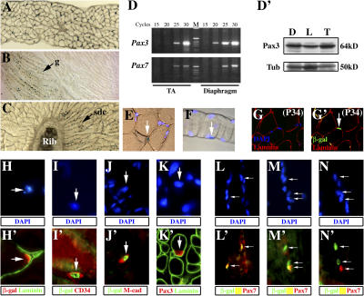
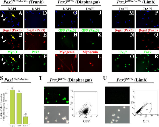
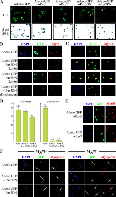
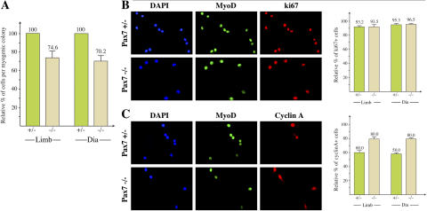
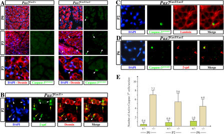
The growth and repair of skeletal muscle after birth depends on satellite cells that are characterized by the expression of Pax7. We show that Pax3, the paralogue of Pax7, is also present in both quiescent and activated satellite cells in many skeletal muscles. Dominant-negative forms of both Pax3 and -7 repress MyoD, but do not interfere with the expression of the other myogenic determination factor, Myf5, which, together with Pax3/7, regulates the myogenic differentiation of these cells. In Pax7 mutants, satellite cells are progressively lost in both Pax3-expressing and -nonexpressing muscles. We show that this is caused by satellite cell death, with effects on the cell cycle. Manipulation of the dominant-negative forms of these factors in satellite cell cultures demonstrates that Pax3 cannot replace the antiapoptotic function of Pax7. These findings underline the importance of cell survival in controlling the stem cell populations of adult tissues and demonstrate a role for upstream factors in this context.


Figures

Figure 1.
Figure 1.
Pax3 expression in muscle satellite cells. (A–C) Expression of Pax3 in different muscles from 3-wk-old Pax3 IRESnLacZ/+ mice, revealed by X-Gal staining. (A) Diaphragm muscle; (B) hindlimb muscles, (g) gracilis muscle; (C) trunk muscles, (sdc) serratus dorsalis caudalis. (D) Semiquantitative RT-PCR of Pax3 and -7 transcripts in adult tibialis anterior (TA) and diaphragm muscle. The number of cycles is indicated on the top of each lane. M, molecular weight markers (see Materials and methods). (D') Pax3 protein is detected by Western blot in protein extracts from different muscles (D, diaphragm; L, hindlimb; T, ventral trunk muscles) in 3-wk-old mice. Tubulin (Tub) expression is shown as a loading control. (E and F) Pax3 IRESnLacZ/+ is expressed in a subset of diaphragm muscle nuclei (arrows) from 3-wk-old mice, as revealed by X-Gal and DAPI staining of a transverse section (E) or isolated fiber (F). (G and G') Detection of Pax3/7-dependent β-gal expression in transverse sections of adult diaphragm muscle from the transgenic line P34, which reports Pax3/7 transcriptional activity. (G) Immunodetection of laminin (red) and DAPI (blue) staining. (G') Coimmunodetection of laminin (red) and β-gal (green). (H–J') Coimmunodetection of β-gal–positive cells in the diaphragm muscle of 3-wk-old Pax3 IRESnLacZ/+ mice with laminin (H', green), CD34 (I', red), or M-cadherin (J', red). As indicated in the figure, β-gal is shown in red in H' and in green in I' and J'. Corresponding DAPI staining is shown (H, I, and J) for each panel. Arrows indicate the labeled satellite cell nuclei. (K) Coimmunodetection of Pax3 (red) and laminin (green, K'). Corresponding DAPI staining is shown, with the labeled nucleus indicated by an arrow (K). (L–N') Coimmunohistochemistry on diaphragm muscle from 3-wk-old Pax3 IRESnLacZ/+ mice for Pax7 (red) or β-gal (green). (L–N) Corresponding DAPI staining is presented with labeled nuclei indicated by arrows for each panel.
Figure 2.
Figure 2.
Pax3 and -7 expression in activated satellite cells. (A–F) Coimmunocytochemistry on primary cultures derived from the trunk muscles of 3-wk-old Pax3 nLacZ/+ mice after 4 d in culture, using DAPI staining (A and D), or an antibody recognizing β-gal (Pax3; B and E, red), MyoD (C, green) or Pax7 (F, green). Whereas β-gal (Pax3) and MyoD are coexpressed in proliferating myoblasts, upon terminal differentiation Pax3 (β-gal) is down-regulated (A–C, arrowheads), and is already lower in some mononucleated MyoD-positive cells (A–C, arrow). (E and F) Most colonies coexpress β-gal (Pax3) and Pax7. (G–L) Detection of GFP and myogenin in primary cultures of satellite cells from the diaphragm of 3-wk-old Pax3GFP/+ mice after 5 d in culture; (G and J) DAPI staining; (H and K) direct detection of GFP (Pax3) fluorescence (green); and (I and L) immunodetection of myogenin (red). (M–R) Coimmunocytochemistry on primary cultures derived from the hindlimb muscles of 3-wk-old Pax3 nLacZ/+ mice after 4 d in culture, showing DAPI staining (M and P) or reaction with an antibody recognizing β-gal (N and Q, red) or Pax7 (O and R, green). Colonies expressing either Pax7 alone (N and O) or Pax3 and -7 (Q and R; ≤20%, see S) were identified. (S) Histograms showing the percentage (%) of β-gal (Pax3)–positive colonies of myogenic cells obtained from the diaphragm (Diaph.), ventral trunk (Trunk), and hindlimb (Limb) muscles of 3-wk-old Pax3 nLacZ/+ mice. Cells were plated at low density to permit the formation of colonies and stained with X-Gal 3–4 d after plating. The results are from three independent experiments after counting ≥100 colonies from triplicate culture plates. (T and U) Muscle satellite cells were isolated by flow cytometry from the lower hindlimb muscles and from the diaphragm of Pax3GFP/+ adult mice as (Pax3) GFP-negative and (Pax3) -positive cells and maintained in culture as proliferating cells for 6 d before analysis of GFP expression by flow cytometry. Direct detection of GFP was performed on living cells under an inverted fluorescence microscope (T and U, top left) with corresponding phase-contrast detection of the cells (T and U, bottom left) and flow cytometry detection of GFP-positive cells (T and U, right, boxed R2 region).
Figure 3.
Figure 3.
The effect of dominant-negative forms of Pax3 and -7 on MyoD and Myf5 expression in satellite cells. (A) Primary cultures prepared from adult diaphragm muscle from the Pax3/7 transcriptional reporter line, P34, were infected with adenoviruses encoding either GFP alone (Adeno-GFP), GFP and Pax3, GFP and Pax7, or GFP with dominant-negative versions of either Pax3 (Pax3DN) or Pax7 (Pax7DN). (top) Infected cells are identified as GFP positive. (bottom) The capacity of Pax3DN or -7DN to abrogate Pax3/7 transcriptional activity is revealed by weaker (arrows) or undetectable X-Gal staining seen in ≥80% of GFP-positive cells with both dominant-negative constructs. (B and C) Coimmunohistochemistry on primary cultures from hindlimb or diaphragm muscles of 3-wk-old wild-type mice infected with adenoviral vectors as in A. DAPI staining and antibodies recognizing either Myf5 (B) or MyoD (C) were used, whereas GFP fluorescence was detected directly. Cells expressing lower levels of PaxDN are indicated with an arrowhead. (D) Quantitation of results for satellite cell cultures infected with an adenovirus-expressing GFP and Pax3DN or GFP and Pax7DN, presented in B and C. Results presented as histograms of the percentage (%) of GFP-positive cells expressing Myf5 or MyoD are taken from two to five independent experiments. (E) Primary cultures of satellite cells isolated from the diaphragm muscle of adult wild-type mice were infected with adenoviruses expressing GFP and Pax3 or -7. Infected cells were identified by direct detection of GFP fluorescence, and MyoD expression was followed by immunodetection. (F) Primary cultures of muscle satellite cells from hindlimb muscles of 3-wk-old Myf5 GFP/+ and Myf5 GFP/GFP mice were infected with adenoviral vectors encoding either GFP alone or GFP and Pax3DN or -7DN. Infected cells are identified by GFP fluorescence, and myogenesis is monitored by immunodetection of myogenin. The threshold of fluorescent GFP detection was set to capture only the strong adenovirus GFP reporter, excluding the weaker expression from the Myf5 GFP allele.
Figure 4.
Figure 4.
Pax3 expression is maintained in Pax7-deficient satellite cells. (A) Western blot analysis of Pax3 expression in the diaphragm of Pax7 LacZ/+ (+/−) or Pax7 LacZ/LacZ (−/−) mice at P3. Tubulin (Tub) expression is shown as a loading control. (B) Coimmunohistochemistry on transverse sections of ventral trunk muscle of Pax7 LacZ/LacZ mice at P2 using DAPI staining and an antibody that recognizes Pax3. Laminin staining shows a Pax3-positive cell (red) present in a satellite cell position. (C) Immunohistochemistry on transverse sections of ventral trunk muscle from Pax7 LacZ/+ or Pax7 LacZ/ LacZ mice at P2 using antibodies recognizing β-gal (green) and laminin (red). Corresponding DAPI staining is indicated. Arrowheads indicate Pax7 (β-gal)–expressing cells, located under the basal lamina. (D) Coimmunocytochemistry on 7-d primary cultures derived from the diaphragm of Pax7 LacZ/+ or Pax7 LacZ/LacZ mice at P10 using antibodies recognizing MyoD and troponin T. (E) Quantification of the number of β-gal–positive satellite cells in trunk, diaphragm (dia), and hindlimb (limb) muscles of Pax7 LacZ/+ (+/−) or Pax7 LacZ/LacZ (−/−) mice, detected per fiber on 10-μm sections from ventral trunk muscle at P2 or P11. (F) Infection of primary cultures from Pax7 LacZ/LacZ mutant mice (Pax7 / ) at P4 with an adenovirus expressing a dominant-negative form of Pax3 (Adeno GFP+Pax3DN) shows elimination of MyoD expression in infected cells (right arrowhead, infected cell; left arrowhead, noninfected cell), whereas virus expressing GFP alone had no effect. Quantification is shown in G. No MyoD-positive myogenic cells are present after Pax3DN infection.
Figure 5.
Figure 5.
Satellite cell proliferation in the Pax7 mutant. (A) Satellite cells from the diaphragm (dia) or hindlimb muscles (limb) of Pax7 lacz/+ (+/−) and Pax7 lacz/lacz (−/−) mice at P4 were plated at low density. After 3 d, when colonies had formed, cells were processed for immunocytochemistry using a MyoD antibody. The number of cells per myogenic colony was determined in three independent experiments for each type of muscle and after counting at least 10 colonies per experiment. Results are expressed as the percentage of cells per myogenic colony from the Pax7 mutant (−/−) with respect to Pax7 heterozygotes (+/−), taken as 100%. (B) Determination of the percentage of cycling cells in normal and mutant myogenic colonies cultured as in A. Cells were costained with antibodies against MyoD and Ki67, which marks all phases of the cell cycle. Examples shown are for myogenic colonies from hindlimb muscles. Quantitation is from two independent experiments for each type of muscle. Results expressed as percentage of Ki67-positive cells per myogenic colony indicate that both mutant and wild type cells proliferate equally well. (C) Determination of the percentage of cyclin A–positive cells per myogenic colony cultured as in A. Cells were costained with antibodies against MyoD and cyclin A, which marks the S and G2 phases of the cell cycle. Examples shown are from hindlimb muscles treated for immunocytochemistry as in B. Results are expressed as percentage of cyclin A–positive cells per myogenic colony.
Figure 6.
Figure 6.
Satellite cell survival in Pax7 mutant mice. (A) Coimmunohistochemistry on transverse sections of ventral trunk muscle of Pax7 LacZ/+ or Pax7 LacZ/LacZ newborn mice at P0 or P3 using DAPI staining or antibodies recognizing desmin (red) or the activated form of caspase-3 (green). Apoptotic cells that are desmin positive are present in muscles from Pax7 mutant mice (arrowheads). (B) Coimmunohistochemistry on transverse sections of ventral trunk muscle of Pax7 LacZ/+ mice at P2 using DAPI staining or antibodies recognizing desmin (red) or β-gal (green). Pax7 (β-gal)-expressing satellite cells are either desmin positive (arrows), which marks activated satellite cells, or desmin negative (arrowheads), indicating quiescent satellite cells. (C and D) Coimmunohistochemistry on transverse sections of ventral trunk muscle of Pax7 LacZ/LacZ mice at P2 (C) or P6 (D) using DAPI staining or antibodies recognizing the activated form of caspase-3 (green), laminin (C, red), or β-gal (D, red) shows that the Pax7 mutant cells located in a satellite cell position are subject to apoptosis. (E) Quantification of apoptotic cells at P0, P2, and P6, based on analysis of sections after coimmunohistochemistry with an antibody to activated caspase-3, and to desmin as a marker of muscle cells. Cells labeled with both antibodies were scored per standard transverse section of trunk and forelimb muscle.
Figure 7.
Figure 7.
The role of Pax7 and -3 in the survival of satellite cells. (A) An example of the data obtained by flow cytometry after infection of cells cultured from hindlimb muscles with a GFP-labeled adenovirus expressing GFP alone or with a dominant-negative form of Pax3 (Pax3DN) or Pax7 (Pax7DN). The percentages indicate the number of GFP-positive cells that are undergoing cell death on the basis of PI uptake (R4) compared with those that survive (R3). (B) A summary of results obtained from the hindlimb on infection with both Pax7DN and Pax3DN, based on four independent experiments with triplicate cultures in each experiment. The numbers shown in the histograms are the percentage of dying cells (R4). Control experiments were performed with adenoviruses expressing only GFP. Numbers in brackets indicate the multiplicity of infection. (C) A summary of results obtained for cells isolated from the diaphragm. The histograms correspond to five independent experiments for the control (5–30) and Pax3DN (5–10), two for Pax3DN (30), and three for Pax7DN (5). Numbers in brackets indicate the multiplicity of infection. (D) The number of MyoD-positive colonies per culture dish obtained after plating the same number of cells from hindlimb or diaphragm muscle of Pax7 LacZ/+ or Pax7 LacZ/LacZ mice at P4.

References

    1. Abrams, J.M., and M.A. White. 2004. Coordination of cell death and the cell cycle: linking proliferation to death through private and communal couplers. Curr. Opin. Cell Biol. 16:634–638. - PubMed
    1. Auerbach, R. 1954. Analysis of the developmental effects of a lethal mutation in the house mouse. J. Exp. Zool. 127:305–329.
    1. Beauchamp, J.R., L. Heslop, D.S. Yu, S. Tajbakhsh, R.G. Kelly, A. Wernig, M.E. Buckingham, T.A. Partridge, and P.S. Zammit. 2000. Expression of CD34 and Myf5 defines the majority of quiescent adult skeletal muscle satellite cells. J. Cell Biol. 151:1221–1234. - PMC - PubMed
    1. Ben-Yair, R., and C. Kalcheim. 2005. Lineage analysis of the avian dermomyotome sheet reveals the existence of single cells with both dermal and muscle progenitor fates. Development. 132:689–701. - PubMed
    1. Bischoff, R., and C. Heintz. 1994. Enhancement of skeletal muscle regeneration. Dev. Dyn. 201:41–54. - PubMed

Publication types

MeSH terms