. 2006 Jan 2;172(1):41-53.
doi: 10.1083/jcb.200509124. Epub 2005 Dec 27.

Coupling of the nucleus and cytoplasm: role of the LINC complex

Affiliations

Coupling of the nucleus and cytoplasm: role of the LINC complex

Melissa Crisp et al. J Cell Biol. .

Abstract

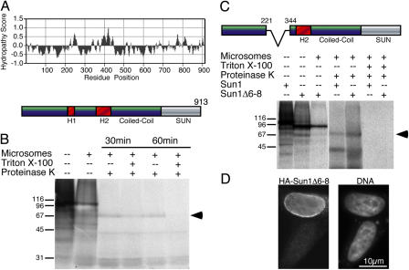
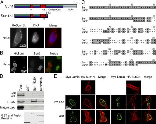
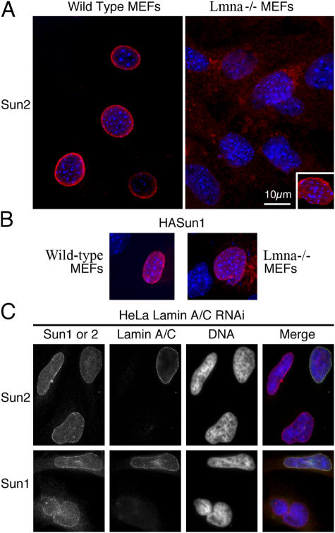
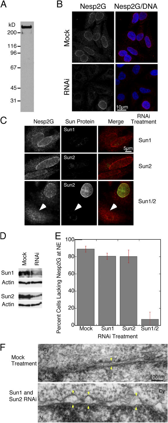
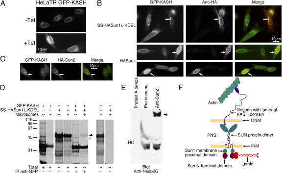
The nuclear envelope defines the barrier between the nucleus and cytoplasm and features inner and outer membranes separated by a perinuclear space (PNS). The inner nuclear membrane contains specific integral proteins that include Sun1 and Sun2. Although the outer nuclear membrane (ONM) is continuous with the endoplasmic reticulum, it is nevertheless enriched in several integral membrane proteins, including nesprin 2 Giant (nesp2G), an 800-kD protein featuring an NH(2)-terminal actin-binding domain. A recent study (Padmakumar, V.C., T. Libotte, W. Lu, H. Zaim, S. Abraham, A.A. Noegel, J. Gotzmann, R. Foisner, and I. Karakesisoglou. 2005. J. Cell Sci. 118:3419-3430) has shown that localization of nesp2G to the ONM is dependent upon an interaction with Sun1. In this study, we confirm and extend these results by demonstrating that both Sun1 and Sun2 contribute to nesp2G localization. Codepletion of both of these proteins in HeLa cells leads to the loss of ONM-associated nesp2G, as does overexpression of the Sun1 lumenal domain. Both treatments result in the expansion of the PNS. These data, together with those of Padmakumar et al. (2005), support a model in which Sun proteins tether nesprins in the ONM via interactions spanning the PNS. In this way, Sun proteins and nesprins form a complex that links the nucleoskeleton and cytoskeleton (the LINC complex).


Figures

Figure 1.
Figure 1.
Sun1 is a ubiquitously expressed NE protein featuring a conserved COOH-terminal SUN domain. (A) ClustalW alignment of the COOH-terminal region of human Sun1–3 and C. elegans Unc-84 reveals the homology of the SUN domains. (B) Northern blot analysis of mRNA from multiple mouse tissues illustrates widespread expression of Sun1. A GAPDH probe was used as a loading control. (C) The NE localizations of Sun1 and 2 were determined by immunofluorescence microscopy using rabbit antibodies raised against recombinant Sun proteins. (D, left) HeLa cells transiently transfected with HA-tagged Sun1 confirm the predominant NE localization. HA-Sun1 was detected using an anti-HA monoclonal antibody. (D, right) Upon overexpression, HA-Sun1 is also detected in the ER. In each case, Sun protein localization is shown in red, whereas DNA, visualized using Höchst dye 33258, appears in blue.
Figure 2.
Figure 2.
Sun1 is a transmembrane protein with a lumenal COOH-terminal domain. (A) Hydropathy plot (Sweet and Eisenberg, 1983) of Sun1 reveals two hydrophobic domains (H1 and H2) upstream of the COOH-terminal coiled-coil and SUN domain. (B) When translated in vitro either in the presence or absence of microsomes, HA-tagged mouse Sun1, labeled with [35S]methionine/cysteine, appears as a ∼100-kD band, as revealed by SDS-PAGE. Subsequent proteinase K digestion of HA-Sun1 that had been translated in the presence of microsomes lead within 30–60 min to the quantitative loss of the full-length HA-Sun1 and the appearance of an ∼65–70-kD protected fragment (arrowhead). Inclusion of Triton X-100 in the digest to permeabilize the microsomes leads to the complete degradation of HA-Sun1 within 60 min. (C) An alternatively spliced isoform of Sun1 (Δ6–8) lacks the first hydrophobic domain (H1). When translated in vitro in either the presence or absence of microsomes, HA-Sun1Δ6–8 appears as a band that is predictably smaller than the full-length protein. However, both full-length HA-Sun1 and HA-Sun1Δ6–8 that were translated in the presence of microsomes and subjected to digestion with proteinase K yield identically sized protected fragments (arrowhead). Inclusion of Triton X-100 in the digestion reaction results in degradation of both proteins. (D) Immunofluorescence microscopy of HeLa cells transfected with HA-Sun1Δ6–8 reveals that the exogenous protein is localized at the NE. In this respect, HA-Sun1Δ6–8 is indistinguishable from full-length HA-Sun1. HA-Sun1Δ6–8 is detected with an anti-HA monoclonal antibody. The corresponding field labeled with Höchst dye to reveal cell nuclei is also shown.
Figure 3.
Figure 3.
The SUN domain of Sun1 is located within the PNS, whereas the NH 2 -terminal domain is exposed to the nucleoplasm. To examine the orientation of Sun1, HA and myc epitope tags were placed at the NH2 and COOH termini, respectively. HeLa cells were transfected with the double-tagged construct and processed for immunofluorescence microscopy after 24 h. After fixation, the cells were permeabilized with 0.003% digitonin and incubated with an anti-epitope tag antibody (mouse monoclonal). Subsequently, the cells were refixed and permeabilized with Triton X-100 to expose the lumenal compartment to a second anti-epitope tag antibody (rabbit polyclonal). Neither epitope tag was significantly detectable after digitonin permeabilization in HeLa cells expressing low to moderate levels of HA-Sun1–myc. After Triton X-100 permeabilization, both myc and HA were readily detected at the NE. In HeLa cells with elevated expression of HA-Sun1–myc, the HA but not myc epitope tag was detected at the ER after digitonin permeabilization. Both myc and HA tags were identifiable after treatment with Triton X-100. These data indicate that the NH2- and COOH-terminal domains of Sun1 reside on opposite sides of the INM, with the COOH-terminal domain located in the PNS.
Figure 4.
Figure 4.
Interactions between the nucleoplasmic domains of Sun1 and 2 with A-type lamins. (A) An HA-Sun1ΔL construct lacking the SUN domain and most of the lumenal coiled-coil localizes to the NE in a manner similar to the full-length protein. Anti-HA labeling is in red, whereas DNA, visualized with Höchst dye, is in blue. (B) Overexpression of HA-Sun1 leads to the loss of endogenous Sun2 in HeLa cells, suggesting that these two proteins share common binding partners. (C) A ClustalW alignment of the NH2-terminal sequences of mouse Sun1 and 2 identifies multiple clusters of homologous amino acids within a region of ∼120 residues. Sun1 exhibits a unique NH2-terminal extension of 50 amino acid residues. (D) The first 222 residues of Sun1 or the first 165 residues of Sun2 (this represents the entire nucleoplasmic domain of the latter) were fused to GST and, with GST alone (Coomassie stain, bottom), were used to pull down 35S-labeled, in vitro translated lamins B1, C, mature A, and full-length (FL) A (Total). Unlike GST alone, Sun1N220 and Sun2N165 pulled down lamins B1, C, and mature A at similar levels. However, Sun1N222 displayed a higher affinity for FL LaA than did Sun2N165. Evidently, Sun1 has a very strong preference for full-length (or pre) lamin A over mature lamin A (or full-length lamins C and B1). (E) An HA tag was inserted at the NH2 terminus of the same nucleoplasmic segments of both Sun1 and 2 (as described in D), and the tagged proteins were expressed in HeLa cells. Both of these exogenous proteins appear enriched in the nucleoplasm (top). As observed by deconvolution microscopy, cotransfection of the myc-tagged full-length lamin A (green) with HA-Sun1N222 or HA-Sun2N165 (red) resulted in the recruitment of both HA-Sun proteins to the NE (middle). Myc–lamin B1, in contrast, fails to produce such an effect. Evidently, the nucleoplasmic domains of Sun1 and 2 can interact with lamin A in vivo.
Figure 5.
Figure 5.
A-type lamin independent retention of SUN domain proteins at the NE. (A) Endogenous Sun2 is frequently, but not always (inset), lost from the NE in Lmna-null MEFs. In wild-type MEFs, Sun2 is always found at the NE. (B) When HA-Sun1 was introduced by transfection into either wild-type or Lmna-null MEFs, it was always found to localize appropriately to the NE. (C) Depletion of HeLa cells of A-type lamins by RNAi had no significant effect on endogenous Sun1 or 2 localization. In the merged images, Sun protein localization is shown in red, A-type lamin localization (lamin A/C) is shown in green, and nuclei are revealed in blue using Höchst dye. The inference is that A-type lamins have, at best, a limited role in the retention of Sun2 at the NE and no significant role at all in the retention of Sun1.
Figure 6.
Figure 6.
The retention of nesp2G at the ONM requires the expression of SUN domain proteins. (A) Western blot of a HeLa lysate fractionated by SDS-PAGE and probed with an affinity-purified antibody raised against the ABD of human nesp2G identifies a very large (>400 kD) protein. (B) Immunofluorescence microscopy of digitonin-permeabilized HeLa cells using the anti-ABD antibody reveals labeling of the cytoplasmic face of the NE. Depletion of nesprin 2 (all splice isoforms) in HeLa cells by RNAi leads to a loss of ONM labeling in the majority of cells (bottom). These data are consistent with the recognition of nesp2G by the anti-ABD antibody. (C) The reduction of either Sun1 or 2 levels by RNAi had only a marginal effect on nesp2G localization. However, the combined depletion of both Sun1 and 2 induced a dramatic loss of nesp2G from the ONM (arrowheads). Overall, we observed an ∼80% decline in the number of cells exhibiting NE-associated nesp2G (E). In total, 100–200 cells were scored for each category in three separate experiments. Error bars represent SEM. (D) Western blot analysis reveals that both Sun1 and 2 RNAi treatments lead to a substantial decline in Sun1 and 2 protein levels. The same blots were probed with an anti-actin antibody to confirm equal loading. (F) Thin section EM of cells subjected to the double (Sun1 and 2) RNAi treatment revealed frequent expansion of the PNS and increased separation of the INM and ONM (arrowheads). No such effect was observed in mock-treated cells. The nuclear interior (N) and cytoplasm (Cy) is indicated in each panel.
Figure 7.
Figure 7.
A soluble form of the Sun1 lumenal domain causes a loss of nesp2G from the ONM. (A) A signal sequence (SS), HA tag, and KDEL motif were added to the NH2 and COOH termini, respectively, of the Sun1 lumenal domain (SS–HA-Sun1L–KDEL). (B) SS–HA-Sun1L–KDEL, when translated in vitro in the presence of microsomes, is completely resistant to digestion by proteinase K. If Triton X-100 is included in the digestion reaction to permeabilize the microsomes, the ∼50-kD SS–HA-Sun1L–KDEL translation product (arrow) is completely degraded. These data demonstrate that the signal sequence is fully functional in directing HA-Sun1L–KDEL to the microsomal lumen. (C) When introduced by transfection into HeLa cells, the SS–HA-Sun1L–KDEL localizes both to the peripheral ER and to the PNS, which is revealed by immunolabeling with an anti-HA monoclonal antibody. Cells expressing SS–HA-Sun1L–KDEL (red in merged images) exhibit a very obvious loss of nesp2G (green in merged images) from the ONM. (D) Thin section EM of HeLa cells expressing SS–HA-Sun1L–KDEL (Transf.) revealed increased separation between the INM and ONM and expansion of the PNS (arrowheads). This effect was not observed in nontransfected cells (NT). The effects of SS–HA-Sun1L–KDEL expression were identical to those observed after codepletion of Sun1 and 2 by RNAi (Fig. 6).
Figure 8.
Figure 8.
The nesp2G KASH domain interacts with the Sun1 lumenal domain. (A) Fluorescence microscopy of HeLa cells expressing a tetracycline-inducible GFP-KASH fusion protein (HeLaTR GFP-KASH). In the absence of tetracycline (−Tet), GFP-KASH is expressed at low levels and is localized exclusively to the NE. After induction with tetracycline for 24 h (+Tet), GFP-KASH is found throughout the peripheral ER as well as the NE. Expression levels of GFP-KASH within the cell population is extremely uniform both before and after induction. (B) Transfection of SS–HA-Sun1–KDEL into the HeLaTR GFP-KASH cells after tetracycline induction resulted in the complete loss of GFP-KASH from the NE. This was accompanied by the formation of cytoplasmic aggregates (arrows, top). Conversely, introduction of full-length HA-Sun1 into these cells resulted in the recruitment of GFP-KASH to the NE (arrows, middle and bottom). (C) HA-Sun2 was found to have a similar effect (arrows). In the merged images in B and C, GFP-KASH is presented in green, whereas HA-Sun is shown in red. (D) To identify an in vitro interaction between KASH and SUN domains, GFP-KASH, SS–HA-Sun1–KDEL, or both proteins were translated in reticulocyte lysate containing [35S]methionine/cysteine in either the presence or absence of microsomes (Totals). Anti-GFP immunoprecipitation of a fraction of each sample revealed the pull-down of SS–HA-Sun1–KDEL by GFP-KASH when both proteins were cotranslated in the presence of microsomes (arrow). A slightly faster migrating band (asterisk) was detected in the absence of microsomes. However, this band originates in the GFP-KASH translation and is unrelated to HA-Sun1L–KDEL. Molecular masses are indicated in kD. (E) Immunoprecipitation of HeLa cell lysates with anti-Sun2 antibodies coprecipitates a very large anti-nesp2G immunoreactive protein (arrow). HC indicates the position of immunoglobulin heavy chains. These data suggest a significant interaction between KASH and Sun proteins that must involve their lumenal domains. These findings allow us to propose a model for the LINC complex (F) in which nuclear components, including lamins, bind to the INM SUN domain proteins. They, in turn, bind to the KASH domain of the actin-associated giant nesprins on the ONM. Thus, the LINC complex establishes a physical connection between the nucleoskeleton and the cytoskeleton.

References

    1. Adam, S.A., R.E. Sterne-Marre, and L. Gerace. 1990. Nuclear protein import in permeabilized mammalian cells requires soluble cytoplasmic factors. J. Cell Biol. 111:807–816. - PMC - PubMed
    1. Apel, E.D., R.M. Lewis, R.M. Grady, and J.R. Sanes. 2000. Syne-1, a dystrophin- and Klarsicht-related protein associated with synaptic nuclei at the neuromuscular junction. J. Biol. Chem. 275:31986–31995. - PubMed
    1. Broers, J.L., E.A. Peeters, H.J. Kuijpers, J. Endert, C.V. Bouten, C.W. Oomens, F.P. Baaijens, and F.C. Ramaekers. 2004. Decreased mechanical stiffness in LMNA−/− cells is caused by defective nucleo-cytoskeletal integrity: implications for the development of laminopathies. Hum. Mol. Genet. 13:2567–2580. - PubMed
    1. Burke, B., and C.L. Stewart. 2002. Life at the edge: the nuclear envelope and human disease. Nat. Rev. Mol. Cell Biol. 3:575–585. - PubMed
    1. Burnette, W.N. 1981. ‘Western blotting’: electrophoretic transfer of proteins from sodium dodecyl sulfate-polyacrylamide gels to unmodified nitrocellulose and radiographic detection with antibody and radioiodinated protein A. Anal. Biochem. 112:195–203. - PubMed

Publication types

MeSH terms