. 2006 Jan;48(1):16-28.
doi: 10.1002/dev.20112.

Spontaneous facial motility in infancy: a 3D kinematic analysis

Affiliations

Spontaneous facial motility in infancy: a 3D kinematic analysis

Jordan R Green et al. Dev Psychobiol. 2006 Jan.

Abstract

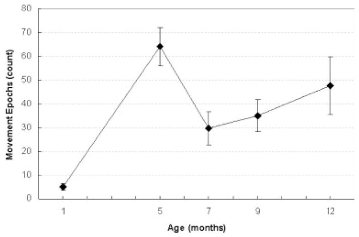
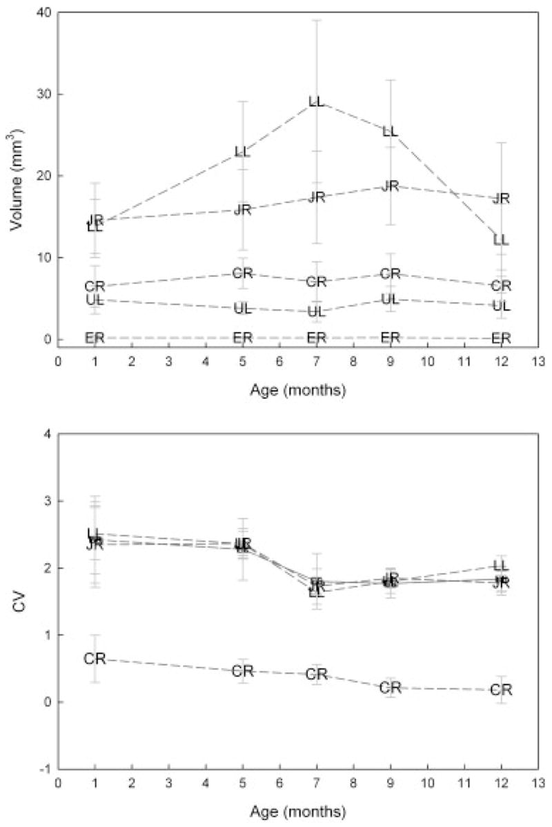
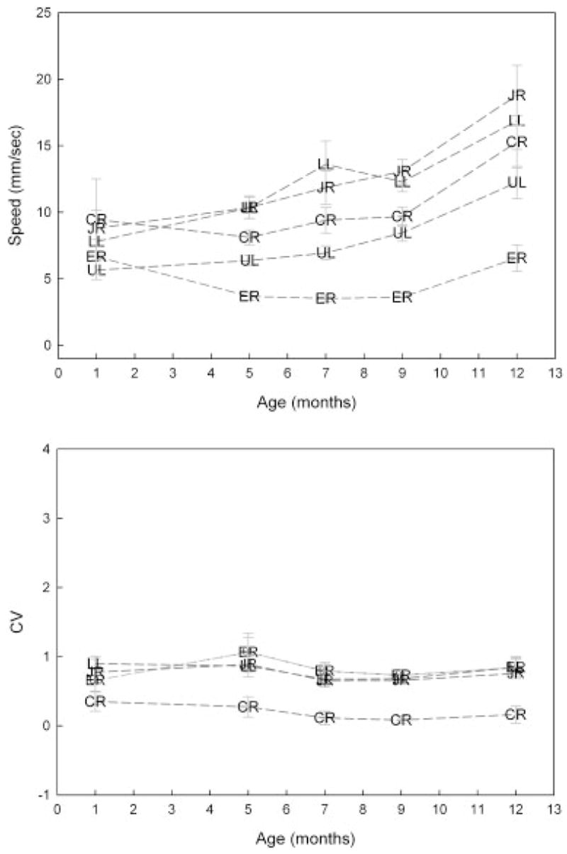
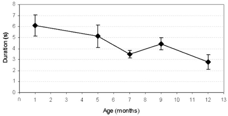
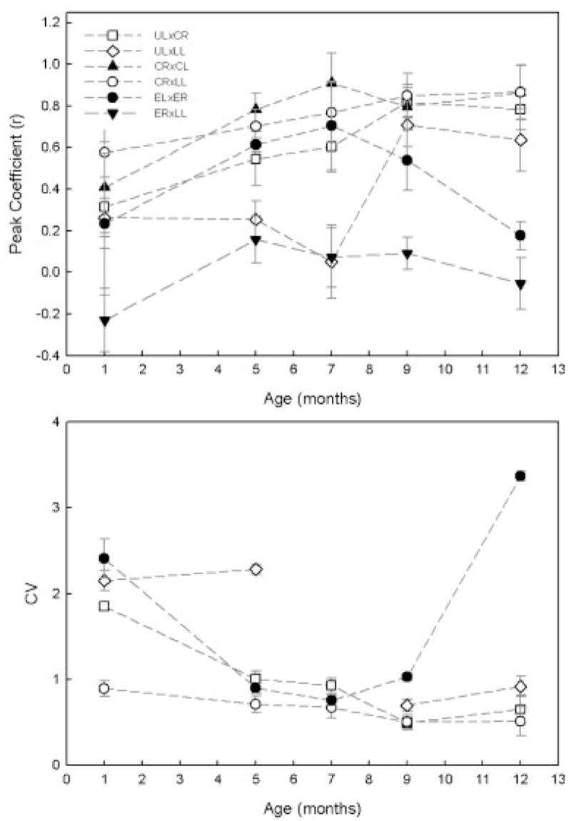
Early spontaneous orofacial movements have rarely been studied experimentally, though the motor experiences gained from these behaviors may influence the development of motor skills emerging for speech. This investigation quantitatively describes developmental changes in silent, spontaneous lip and jaw movements from 1 to 12 months of age using optically based 3D motion capture technology. Twenty-nine typically developing infants at five ages (1, 5, 7, 9, and 12 months) were studied cross-sectionally. Infants exhibited spontaneous facial movements at all ages studied. Several age-related changes were detected in lip and jaw kinematics: the occurrence of spontaneous movements increased, movement speed increased, the duration of movement epochs decreased and movement coupling among different facial regions increased. Additionally, evidence for stereotypic movements was not strong. The present findings suggest that, during the first year of life, early spontaneous facial movements undergo significant developmental change in the direction of skill development for speech.


Figures

FIGURE 1
FIGURE 1
Left panel: A child fitted with the facial marker array. Right panel: 3D facial model reconstruction. For movement tracking, flat, circular reflective markers (32 mm in diameter) were placed on selected facial landmarks. The four reference markers placed on the forehead were used to correct for head movement that would otherwise be included in the facial movement signals. These four markers translate the origin to the head marker array and align the axis to the lines defined by these markers. Thus, all the facial kinematic data were expressed relative to the coordinate system defined by the four head makers. ER, right eyebrow; EL, left eyebrow; UL, upper lip; CR, right oral commissure; CL, left oral commissure; LL, lower lip; JR, chin.
FIGURE 2
FIGURE 2
An example jaw kinematic trace recorded during a spontaneous movement epoch. The 3D distance time-history is based on the markers Euclidean distance from the head-based origin. Speed time-histories were derived by computing the first-order derivative of each marker’s 3D distance time-history.
FIGURE 3
FIGURE 3
An example of the 3D movement space analysis performed on the jaw motion path obtained from a 7-month-old infant during a spontaneous movement.
FIGURE 4
FIGURE 4
The average number of spontaneous movement epoch recorded at each age.
FIGURE 5
FIGURE 5
Top panel: Age-related changes in movement space for selected facial markers. Standard error bars represent across subject variation in each age group. Bottom panel: Age-related changes in the within variability in movement space. ER, right eyebrow; UL, upper lip; CR, right oral commissure; LL, lower lip; JR, chin.
FIGURE 6
FIGURE 6
Top panel: Age-related changes in average movement speed for selected facial markers. Standard error bars represent across subject variation in each age group. Bottom panel: Age-related changes in the within variability in average movement speed. ER, right eyebrow; UL, upper lip; CR, right oral commissure; LL, lower lip; JR, chin.
FIGURE 7
FIGURE 7
Age-related changes in movement epoch duration. Standard error bars represent across subject variation in each age group.
FIGURE 8
FIGURE 8
Top panel: Age-related changes in movement coupling between selected facial markers. Standard error bars represent across subject variation in each age group. Bottom panel: Age-related changes in the within variability in movement coupling. The CV values for ER3LL at all ages studied and for UL3LL at 7 months were very large (e.g., 11.5) and therefore not displayed. ER, right eyebrow; EL, left eyebrow; UL, upper lip; CR, right oral commissure; CL, left oral commissure; LL, lower lip; JR, chin.

References

    1. Barlow SM, Finan DS, Rowland SG. Mechanically evoked perioral reflexes in infants. Brain Research. 1992;599:158–160. - PubMed
    1. Bekoff A. Ontogeny of chicken motor behaviors: Evidence for multiuse limb pattern generating circuitry. In: Grillner S, Stein PSG, Stuart DG, Forssberg H, Herman RM, editors. Neurobiology of vertebrate locomotion. Hampshire, England: Macmillan Press; 1986. pp. 433–453.
    1. Bekoff A. Spontaneous embryonic motility: An enduring legacy. International Journal of Developmental Neuroscience. 2001;19:155–160. - PubMed
    1. Birnholz JC, Stephens JC, Faria M. Fetal movement patterns: A possible means of defining neurologic developmental milestones in utero. American Journal of Roentgenology. 1978;130:537–540. - PubMed
    1. Coghill GE. Anatomy and the problem of behaviour. New York: Cambridge University Press; 1929.

Publication types