. 2006 Jan 10;103(2):437-42.
doi: 10.1073/pnas.0510012103. Epub 2005 Dec 28.

The E7 proteins of low- and high-risk human papillomaviruses share the ability to target the pRB family member p130 for degradation

Affiliations

The E7 proteins of low- and high-risk human papillomaviruses share the ability to target the pRB family member p130 for degradation

Benyue Zhang et al. Proc Natl Acad Sci U S A. .

Abstract

High-risk human papillomaviruses (HPVs) (e.g., HPV-16) cause anogenital and head and neck cancers, and low-risk HPVs (e.g., HPV-6) cause benign hyperproliferative disease. The E7 protein of HPV-16 binds all retinoblastoma tumor suppressor protein (pRB) family members with higher affinity than HPV-6E7. The HPV-16 E7 protein has been reported to target pRB family members for degradation and to immortalize cells. Here we tested the hypothesis that the low-risk E7 protein has an intrinsic ability to decrease expression of pRB family members. First, we introduced a high-affinity pRB-binding site into HPV-6 E7 (6E7G22D) and showed that, in human foreskin keratinocytes, HPV-6 E7G22D decreased the level of pRB protein but not pRB mRNA. Second, we analyzed the ability of wild-type HPV-6 E7 to destabilize the other pRB family members, p107 and p130. HPV-6 E7, like HPV-16 E7, decreased the level of p130 protein. This decrease was inhibited by MG132, a proteasome inhibitor. Binding of HPV-6 E7 to p130 was necessary but not sufficient to decrease the level of p130. Furthermore, the destabilization of p130 correlated with a decrease in the expression of involucrin, a differentiation marker. We suggest that the shared activity of HPV-16 E7 and HPV-6 E7 to destabilize p130 and decrease or delay differentiation may be related to the role of E7 in the HPV life cycle. The added ability of HPV-16 E7 to regulate pRB and p107 may be related to oncogenic activity.


Figures
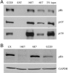
Fig. 1.
Fig. 1.
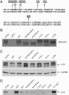
Characterization of HPV-6 E7G22D with respect to binding of pRB family members and degradation of pRB. (A) The ability of HPV-6, HPV-16, and HPV-6 E7G22D to bind pRB family members. Jurkat nuclear extracts were incubated with GST, GST-16E7, GST-6E7, or GST-6E7G22D, and bound proteins were detected after separation by SDS/PAGE and transfer to nitrocellulose membrane. The membrane was probed with antibodies to pRB, p107, and p130. Ponceau red staining of the membrane before probing indicated that similar levels of GST or GST fusion proteins were present in the precipitate (data not shown). The average ± SD of bound pRB in three independent experiments, corrected for Ponceau red and relative to GST16E7 (set to 1.0), was 1.22 ± 0.15 (G22D) and 0.35 ± 0.06 (6E7); the average ± SD of bound p107 was 0.73 ± 0.08 (G22D) and 0.36 ± 0.01 (6E7); the average of bound p130 was 0.42 ± 0.07 (G22D) and 0.23 ± 0.05 (6E7). (B) Immunoblot of pRB in the presence and absence of E7. HFKs were infected with control retrovirus LXSN, L(16E7)SN, or L(6E7)SN and grown in C-K-SFM. Whole-cell lysates were analyzed by immunoblot by using antibodies to pRB and GAPDH, the latter as a loading control. The average ± SD of pRB in three independent experiments, corrected for GAPDH and relative to LXSN, was 0.24 ± 0.14 (16E7), 0.89 ± 0.32 (6E7), and 0.50 ± 0.19 (G22D).
Fig. 2.
Fig. 2.
The effect of HPV E7 proteins on p130 expression in undifferentiated and differentiated conditions. (A) Immunoblot of pRB family members in the presence and absence of E7 and in different growth conditions. HFKs were infected with control retrovirus LXSN, L(16E7)SN, or L(6E7)SN and grown in C-K-SFM or C-K-SFM containing 2 mM CaCl2 for 48 h. Whole-cell lysates were analyzed by immunoblot by using antibodies to pRB, p107, p130, and GAPDH. The average ± SD in three independent experiments, corrected for GAPDH and relative to LXSN in C-K-SFM, of pRB was 0.21 ± 0.09 (16E7) and 1.14 ± 0.20 (6E7) in C-K-SFM and 0.32 ± 0.13 (LXSN), 0.01 ± 0.02 (16E7), and 0.22 ± 0.08 (6E7) in CaCl2; the average ± SD of p107 was 0.27 ± 0.18 (16E7) and 1.14 ± 0.19 (6E7) in C-K-SFM and 0.25 ± 0.09 (LXSN), 0.26 ± 0.09 (16E7), and 0.27 ± 0.12 (6E7) in CaCl2; the average ± SD of p130 was 0.35 ± 0.04 (16E7) and 0.36 ± 0.08 (6E7) in C-K-SFM and 1.21 ± 0.06 (LXSN), 0.35 ± 0.05 (16E7), and 0.42 ± 0.04 (6E7) in CaCl2. *, Unknown protein cross-reacting with anti-p130. (B) Analysis of p130 mRNA levels. HFKs were infected and grown as in A, and total RNA was analyzed by RT-PCR with primers amplifying p130 and GAPDH. The results shown are for 26 cycles; similar results were obtained at 22 and 30 cycles. Data are representative of three independent experiments.
Fig. 3.
Fig. 3.
The half life of p130 in the presence and absence of E7 protein and in the presence and absence of a proteasome inhibitor. (A) Decreased half-life of p130 in the presence of HPV E7 protein. HFKs were infected with control retrovirus LXSN, L(16E7)SN, or L(6E7)SN and treated with 0.25 mM cycloheximide for the indicated times. Immunoblots of whole-cell lysates are shown (Left), and densitometry is plotted for the average ± SD of three independent experiments (Right). (B) Inhibition of p130 degradation by MG132. HFKs were infected as in A and treated with 0.25 mM cycloheximide plus 50 μM MG132 for the indicated times. Immunoblots of whole-cell lysates are shown (Left), and densitometry is plotted for the average ± SD of three independent experiments (Right).
Fig. 4.
Fig. 4.
Construction of HPV-6 E7 mutants and their characterization with respect to p130 binding and degradation. (A) Map of HPV-6 E7 mutants. (B) Stability of mutants. COS-7 cells were transfected with pSG5, pSG5(6E7), pSG5(H2AR4AH5A), pSG5(K9AD10A), pSG5(C25A), pSG5(D31A), and pSG5(L67R). Forty-eight hours after transfection, the cells were labeled with 500 μCi of 35S-labeled methionine and cysteine and lysed, and HPV-6 E7 was immunoprecipitated by a polyclonal antibody (anti-6E7). The immunoprecipitates were separated on 15% SDS/PAGE gel and exposed to x-ray film. (C) Amino acids required for destabilization of p130. HFKs were infected with LXSN, L(16E7)SN, L(6E7)SN, or retroviruses encoding the HPV-6 E7 mutants. Whole-cell lysates from transduced cells grown in C-K-SFM were analyzed by Western blot with antibodies to p130. The average ± SD of p130 in three independent experiments, corrected for GAPDH and relative to LXSN, was 0.33 ± 0.06 (16E7), 0.35 ± 0.06 (6E7), 1.03 ± 0.05 (H2AR4AH5A), 0.43 ± 0.06 (K9AD10A), 0.79 ± 0.15 (C25A), 0.37 ± 0.07 (D31A), and 0.89 ± 0.32 (L67R). (D) E7 amino acids required for binding to p130. GST pull-downs were conducted as in Fig. 1 with GST, GST-16E7, GST-6E7H2AR4AH5A, GST-6E7K9AD10A, GST-6E7C25A, GST-6E7D31A, and GST-6E7L67R, and the immunoblots were probed with antibodies to p130. Ponceau red staining indicated equal amounts of GST and GST fusion proteins in the precipitate (data not shown). The average ± SD of bound p130 in three independent experiments, corrected for Ponceau red and relative to GST6E7, was 6.86 ± 2.40 (16E7), 1.23 ± 0.76 (H2AR4AH5A), 0.51 ± 0.18 (K9AD10A), 0.16 ± 0.09 (C25A), 1.12 ± 0.12 (D31A), and 0.71 ± 0.05 (L67R).
Fig. 5.
Fig. 5.
HPV E7-mediated decreased/delayed keratinocyte differentiation and correlation with the ability of HPV-6 E7 to destabilize p130. (A) Effect of HPV-16 E7 and HPV-6 E7 on differentiation. HFKs were transduced and grown as described in Fig. 1. Whole-cell lysates were analyzed on Western blots by using antibodies to involucrin, keratin 10, and GAPDH. The average ± SD of involucrin in three independent experiments, corrected for GAPDH, are as follows: relative to LXSN in C-K-SFM, 0.54 ± 0.17 (16E7) and 0.35 ± 0.18 (6E7); relative to LXSN in CaCl2 for 24 h, 0.46 ± 0.11 (16E7) and 0.45 ± 0.12 (6E7); relative to LXSN in CaCl2 for 48 h, 0.63 ± 0.15 (16E7) and 0.54 ± 0.19 (6E7). The average ± SD of keratin 10 in three independent experiments, corrected for GAPDH and relative to LXSN in CaCl2 for 48 h, was 0.04 ± 0.00 (16E7) and 0.15 ± 0.05 (6E7). (B) Effect of HPV-6 E7 and HPV-6 E7 mutants on differentiation. HFKs were infected and grown as in Fig. 1, and immunoblots were performed on whole-cell extracts by using antibodies to p130, involucrin, and GAPDH. The average ± SD of p130 in three independent experiments, corrected for GAPDH, was as follows: relative to LXSN in C-K-SFM, 0.31 ± 0.08 (6E7); relative to LXSN in CaCl2, 0.43 ± 0.02 (6E7), 1.08 ± 0.10 (H2AR4AH5A), 0.52 ± 0.13 (K9AD10A), 0.86 ± 0.09 (C25A), 0.48 ± 0.08 (D31A), and 0.88 ± 0.10 (L67R); the average ± SD of involucrin relative to LXSN in CaCl2 was 0.38 ± 0.19 (6E7), 0.89 ± 0.03 (H2AR4AH5A), 0.51 ± 0.14 (K9AD10A), 0.88 ± 0.14 (C25A), 0.43 ± 0.09 (D31A), and 0.79 ± 0.01 (L67R).

Similar articles

Cited by

References

    1. zur Hausen, H. (2002) Nat. Rev. Cancer 2, 342-350. - PubMed
    1. Longworth, M. S. & Laimins, L. A. (2004) Microbiol. Mol. Biol. Rev. 68, 362-372. - PMC - PubMed
    1. Cheng, S., Schmidt-Grimminger, D. C., Murant, T., Broker, T. R. & Chow, L. T. (1995) Genes Dev. 9, 2335-2349. - PubMed
    1. Thomas, J. T., Hubert, W. G., Ruesch, M. N. & Laimins, L. A. (1999) Proc. Natl. Acad. Sci. USA 96, 8449-8454. - PMC - PubMed
    1. Flores, E. R., Allen-Hoffmann, B. L., Lee, D. & Lambert, P. F. (2000) J. Virol. 74, 6622-6631. - PMC - PubMed

Publication types

Substances

LinkOut - more resources