. 2006 Jan 10;103(2):389-94.
doi: 10.1073/pnas.0509978103. Epub 2005 Dec 28.

Transcriptional response of steady-state yeast cultures to transient perturbations in carbon source

Affiliations

Transcriptional response of steady-state yeast cultures to transient perturbations in carbon source

Michal Ronen et al. Proc Natl Acad Sci U S A. .

Abstract

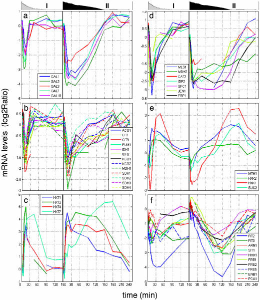
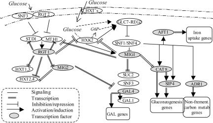
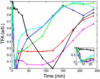
To understand the dynamics of transcriptional response to changing environments, well defined, easily controlled, and short-term perturbation experiments were undertaken. We subjected steady-state cultures of Saccharomyces cerevisiae in chemostats growing on limiting galactose to two different size pulses of glucose, well known to be a preferred carbon source. Although these pulses were not large enough to change growth rates or cell size, approximately 25% of the genes changed their expression at least 2-fold. Using DNA microarrays to estimate mRNA abundance, we found a number of distinguishable patterns of transcriptional response among the many genes whose expression changed. Many of these genes were already known to be regulated by particular transcription factors; we estimated five potentially relevant transcription factor activities from the observed changes in gene expression (i.e., Mig1p, Gal4p, Cat8p, Rgt1p, Adr1p, and Rcs1p). With these estimates, for two regulatory circuits involving interaction among multiple regulators we could generate dynamical models that quantitatively account for the observed transcriptional responses to the transient perturbations.


Figures

Fig. 1.
Fig. 1.
Glucose (○) and ethanol (▵) concentrations observed after the 0.2 g/liter (a) and 2.0 g/liter (b) pulses of glucose. The glucose concentrations predicted solely from dilution in the chemostat are shown by the solid line.
Fig. 2.
Fig. 2.
Gene expression patterns of GAL genes (a), tricarboxylic acid cycle genes (b), glucose transporters (c), gluconeogenesis (d), genes with bidirectional response (e), and iron homeostasis genes (f). The left side of each plot shows the low-glucose pulse, and the right side shows the high-glucose pulse. The graphs above the panels are area plots of glucose concentration: low-glucose (0.2 g/liter) pulses (I) and high-glucose (2.0 g/liter) pulses (II).
Fig. 3.
Fig. 3.
Mechanism of glucose repression and induction. Glucose is transported into the cell by Hxt transporters with diverse affinities. Intracellular glucose is converted to glucose-6-phosphate primarily by Hxk2p and then fermented to ethanol and CO2 (5). Snf1p protein kinase is one of the main players in glucose repression and the induction pathway; it regulates the activities of TFs of glucose repression genes (Mig1p) and gluconeogenesis (Cat8p-Sip4p and Adr1p). In the presence of glucose, Snf1 is deactivated by the phosphatase, Glc7p-Reg1p, in a Hxk2p-dependent manner (15). Once inhibition by Snf1p is released, Mig1p is phosphorylated, enters the nucleus, and represses the expression of alternative carbon source utilization genes (e.g., GAL4, SUC2, SNF3, and gluconeogenesis TF genes CAT8 and SIP5) (5). Iron uptake and transport genes are induced throughout Snf1p/Snf4p and the TF Rcs1p, independent of iron starvation (22). A separate sensing pathway mediates glucose induction of mainly glucose transporter genes. Extracellular glucose binds to glucose receptors Snf3p or Rgt2p (with high and low affinities, respectively) to generate a signal that inactivates the transcriptional receptor Rgt1p. This signal induces the HXT genes, MIG2, and its own activators, STD1 and MTH1 (29, 16). The glucose signal inhibits Rgt1p-mediated expression by stimulating the degradation of Mth1p and Std1p (28). The two pathways, Snf1-Mig1 and Rgt1, are interconnected in both protein signaling and transcriptional levels mainly throughout Mth1p and Hxk2p, which are regulated by both Mig1p and Rgt1p (18, 30) and have a signaling role in the pathways. In addition, Mig1p regulates one of the sensors of the Rgt1 pathway, Snf3p, where Rgt1p regulates the transporters that bring the Snf1 pathway into action. Another glucose sensing and signaling mechanism, not shown here, is responsible for activation of ribosomal and glycolysis genes and repression of glycogen, trehalose, and stress genes through Gpr1p, cAMP, and PKA (4).
Fig. 4.
Fig. 4.
The estimated TFAs with the 2 g/liter glucose pulse, 1-Mig1 (blue), Gal4 (green), Cat8 (red), Rgt1 (pink), Adr1 (cyan), and Rcs1 (black). (Inset) A zoom-in to the initial time points.
Fig. 5.
Fig. 5.
Bidirectional response model. (a) Mig1-Rgt1 circuits. (b) The estimated TFA during derepression. Blue, Mig1p; pink, Rgt1p; solid line, high pulse; dashed line, low pulse. (Inset) The combined factors pattern of gene activation. (c) The expression of MTH1 (green) and HXK2 (red) genes, measured (solid line) and model-predicted (dashed line)
Fig. 6.
Fig. 6.
Lasting response. (a) Mig1-Cat8 circuit. (b) The estimated active TFs during derepression after a high-glucose pulse. Blue, Mig1p; red, Cat8p; black solid line, Rcs1p; black dashed line, predicted Snf1p activity.

References

    1. Gasch, A. P., Spellman, P. T., Kao, C. M., Carmel-Harel, O., Eisen, M. B., Storz, G., Botstein, D. & Brown, P. O. (2000) Mol. Biol. Cell 11, 4241-4257. - PMC - PubMed
    1. Hughes, T. R., Marton, M. J., Jones, A. R., Roberts, C. J., Stoughton, R., Armour, C. D., Bennett, H. A., Coffey, E., Dai, H., He, Y. D., et al. (2000) Cell 102, 109-126. - PubMed
    1. Ideker, T., Thorsson, V., Ranish, J. A., Christmas, R., Buhler, J., Eng, J. K., Bumgarner, R., Goodlett, D. R., Aebersold, R. & Hood, L. (2001) Science 292, 929-934. - PubMed
    1. Gelade, R., Van de Velde, S., Van Dijck, P. & Thevelein, J. M. (2003) Genome Biol. 4, 233. - PMC - PubMed
    1. Johnston, M. (1999) Trends Genet. 15, 29-33. - PubMed

Publication types

MeSH terms