

. 2006 Jan 1;34(Database issue):D231-4.
doi: 10.1093/nar/gkj062.
SitesBase: a database for structure-based protein-ligand binding site comparisons
Affiliations
- PMID: 16381853
- PMCID: PMC1347425
- DOI: 10.1093/nar/gkj062
Item in Clipboard
SitesBase: a database for structure-based protein-ligand binding site comparisons
Nucleic Acids Res.
.
Abstract
There are many components which govern the function of a protein within a cell. Here, we focus on the molecular recognition of small molecules and the prediction of common recognition by similarity between protein-ligand binding sites. SitesBase is an easily accessible database which is simple to use and holds information about structural similarities between known ligand binding sites found in the Protein Data Bank. These similarities are presented to the wider community enabling full analysis of molecular recognition and potentially protein structure-function relationships. SitesBase is accessible at http://www.bioinformatics.leeds.ac.uk/sb.
Figures
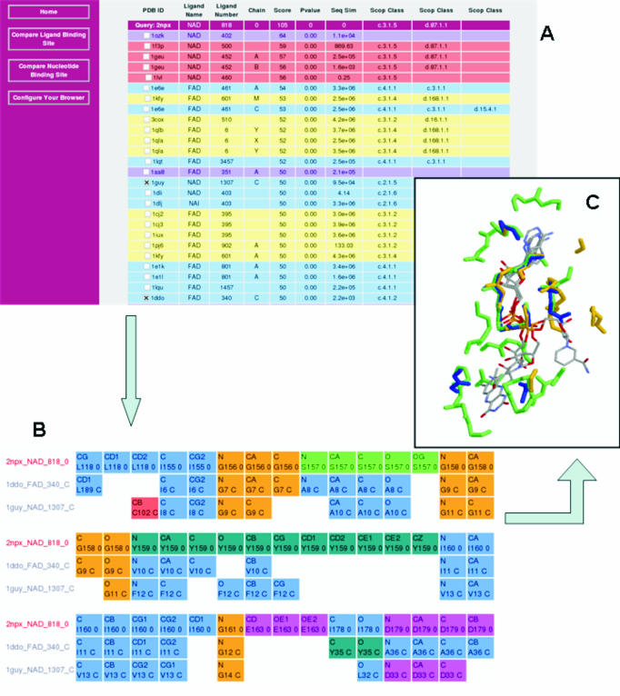
Collection of screen shots from SitesBase showing results of a search (A) with query 2npx (NADH peroxidase). This binding site has two SCOP classes attributed. The primary SCOP class is c.3.1.5 the NAD/FAD reductase family with an NAD/FAD binding domain fold. The SCOP and PDB codes are linked to the SCOP and PDBsum databases, respectively. Four of the listed hits are family relatives of the query (pink), ten are superfamily relatives (yellow) and ten are fold relatives (blue). There are also two high scoring hits which are yet to be classified in the SCOP database (purple). Note: in this illustration some high scoring hits have been removed in order to show more distantly related proteins. (B) Gives a multiple alignment of two selected hits with the same SCOP class (i.e. with different folds) as the query and clearly shows the sequence conserved GXGXXG dinucleotide binding motifs. (C) Illustrates these three protein sites (2npx, blue; 1guy, yellow; and 1ddo, green) superimposed in 3D.
Similar articles
-
New opportunities for protease ligand-binding site comparisons using SitesBase.Biochem Soc Trans. 2007 Jun;35(Pt 3):561-5. doi: 10.1042/BST0350561. Biochem Soc Trans. 2007. PMID: 17511652
-
A searchable database for comparing protein-ligand binding sites for the analysis of structure-function relationships.J Chem Inf Model. 2006 Mar-Apr;46(2):736-42. doi: 10.1021/ci050359c. J Chem Inf Model. 2006. PMID: 16563004
-
Fold independent structural comparisons of protein-ligand binding sites for exploring functional relationships.J Mol Biol. 2006 Feb 3;355(5):1112-24. doi: 10.1016/j.jmb.2005.11.044. Epub 2005 Dec 1. J Mol Biol. 2006. PMID: 16359705
-
The use of protein-ligand interaction fingerprints in docking.Curr Opin Drug Discov Devel. 2008 May;11(3):356-64. Curr Opin Drug Discov Devel. 2008. PMID: 18428089 Review.
-
Protein structure databases with new web services for structural biology and biomedical research.Brief Bioinform. 2008 Jul;9(4):276-85. doi: 10.1093/bib/bbn015. Epub 2008 Apr 22. Brief Bioinform. 2008. PMID: 18430752 Review.
Cited by
-
SitEx: a computer system for analysis of projections of protein functional sites on eukaryotic genes.Nucleic Acids Res. 2012 Jan;40(Database issue):D278-83. doi: 10.1093/nar/gkr1187. Epub 2011 Dec 1. Nucleic Acids Res. 2012. PMID: 22139920 Free PMC article.
-
PESDserv: a server for high-throughput comparison of protein binding site surfaces.Bioinformatics. 2010 Aug 1;26(15):1913-4. doi: 10.1093/bioinformatics/btq288. Epub 2010 Jun 10. Bioinformatics. 2010. PMID: 20538727 Free PMC article.
-
Ligand binding site detection by local structure alignment and its performance complementarity.J Chem Inf Model. 2013 Sep 23;53(9):2462-70. doi: 10.1021/ci4003602. Epub 2013 Sep 4. J Chem Inf Model. 2013. PMID: 23957286 Free PMC article.
-
RNA-protein distance patterns in ribosomes reveal the mechanism of translational attenuation.Sci China Life Sci. 2014 Nov;57(11):1131-9. doi: 10.1007/s11427-014-4753-8. Epub 2014 Oct 18. Sci China Life Sci. 2014. PMID: 25326828 Free PMC article.
-
PoSSuM: a database of similar protein-ligand binding and putative pockets.Nucleic Acids Res. 2012 Jan;40(Database issue):D541-8. doi: 10.1093/nar/gkr1130. Epub 2011 Dec 1. Nucleic Acids Res. 2012. PMID: 22135290 Free PMC article.
References
-
- Laskowski R.A., Hutchinson E.G., Michie A.D., Wallace A.C., Jones M.L., Thornton J.M. PDBsum: a Web-based database of summaries and analyses of all PDB structures. Trends Biochem. Sci. 1997;22:488–490. - PubMed
-
- Murzin A.G., Brenner S.E., Hubbard T., Chothia C. SCOP: a structural classification of proteins database for the investigation of sequences and structures. J. Mol. Biol. 1995;247:536–540. - PubMed
-
- Artymiuk P.J., Poirrette A.R., Grindley H.M., Rice D.W., Willett P. A graph-theoretic approach to the identification of three-dimensional patterns of amino acid side-chains in protein structures. J. Mol. Biol. 1994;243:327–344. - PubMed
-
- Binkowski T.A., Adamian L., Liang J. Inferring functional relationships of proteins from local sequence and spatial surface patterns. J. Mol. Biol. 2003;332:505–526. - PubMed
