. 2006 Jan;26(2):535-47.
doi: 10.1128/MCB.26.2.535-547.2006.

Regulation of sexual dimorphism: mutational and chemogenetic analysis of the doublesex DM domain

Affiliations

Regulation of sexual dimorphism: mutational and chemogenetic analysis of the doublesex DM domain

Wei Zhang et al. Mol Cell Biol. 2006 Jan.

Abstract

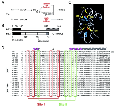
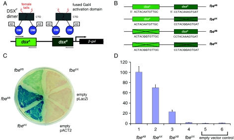
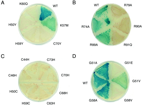
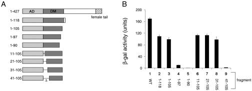
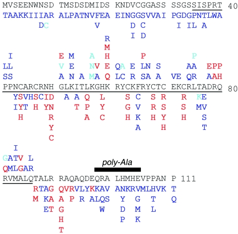
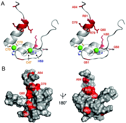
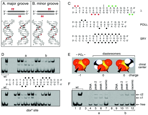
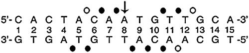
Doublesex (dsx) is a transcription factor in Drosophila that regulates somatic sexual differentiation. Male- and female-specific splicing isoforms of DSX share a novel DNA-binding domain, designated the DM motif. Broadly conserved among metazoan sex-determining factors, the DM domain contains a nonclassical zinc module and binds in the DNA minor groove. Here, we characterize the DM motif by site-directed and random mutagenesis using a yeast one-hybrid (Y1H) system and extend this analysis by chemogenetic complementation in vitro. The Y1H system is based on a sex-specific Drosophila enhancer element and validated through studies of intersexual dsx mutations. We demonstrate that the eight motif-specific histidines and cysteines engaged in zinc coordination are each critical and cannot be interchanged; folding also requires conserved aliphatic side chains in the hydrophobic core. Mutations that impair DNA binding tend to occur at conserved positions, whereas neutral substitutions occur at nonconserved sites. Evidence for a specific salt bridge between a conserved lysine and the DNA backbone is obtained through the synthesis of nonstandard protein and DNA analogs. Together, these results provide molecular links between the structure of the DM domain and its function in the regulation of sexual dimorphism.


Figures

FIG. 1.
FIG. 1.
Sex-determining hierarchy of D. melanogaster and DM motif. (A) X-to-autosome ratio regulates a sex-specific RNA-splicing cascade (50). Not shown are Intersex (IX), a putative transcriptional coactivator that acts with DSXF to promote female development, and a divergent fruitless branch downstream of tra factors. (B) Domain organization of DSX isoforms N-terminal DM and C-terminal dimerization domains. DSXM and DSXF differ at the extreme C terminus (cross-hatched and gray regions). (C) Ribbon model of DM domain with intertwined CCHC and HCCC Zn2+-binding sites (DSX residues 41 to 81) (68). (D) Metazoan DM sequences. Cysteines and histidines that coordinate Zn2+ are aligned (red and green boxes). Stable α-helical elements are highlighted in magenta; the nascent helical tail is shown in gray. The arrow highlights residue K60, conserved on the surface of the Zn module and proposed to contact DNA.
FIG. 2.
FIG. 2.
Design and validation of Y1H system. (A) Specific DSX-DNA recognition regulates the expression of lacZ via fusion protein containing an N-terminal Gal4 AD and a C-terminal DSX domain. A 48-bp Drosophila promoter fragment derived from fbe was inserted upstream of lacZ to provide DSX-binding sites dsxA and dsxB. CTD, C-terminal dimerization domain of DSX. (B) Wild-type DNA-binding sites and inactive variants fbeAB, containing two wild-type sites and control elements (respectively designated fbeAX, fbeXB, and fbeXX) in which one, the other, or both sites are inactivated by twin A→G transitions at center of target sites (bold). (C) Colonies on an X-gal indicator plate in the presence of wild-type or variant enhancer elements. (D) Histogram describing β-galactosidase activity in the presence of wild-type or variant DNA sites (columns 1 to 4), empty reporter vector (column 5) or reporter (column 6) plasmid lacking fbe sites.
FIG. 3.
FIG. 3.
Y1H analysis of variant DM domains. (A) Intersexual mutations H50Y, H59Y, and C70Y inactivate DSX as does K60Q. K57M is light blue, which is consistent with partial activity of isosteric norleucine analog (52). (B) Alanine substitutions previously shown to significantly impair specific DNA binding (R79A and R90A) (52) impair β-galactosidase expression (white colonies), whereas substitutions compatible with high-affinity DNA binding (R74A and R99A) are associated with wild-type β-galactosidase expression (blue). Intersexual mutation R91Q (25) is white. (C) Interchange of histidine and cysteine within the metal-binding sites in each case impairs β-galactosidase expression (white colonies). (D) Variable phenotypes G51A and G58A (blue), G51V (light blue), and G51E and G58V (white). G51 and G58 participate in turns.
FIG. 4.
FIG. 4.
Deletion analysis of DM domain. (A) Set of DSX fragments of different lengths employed in AD fusion proteins (Fig. 2A). The DM domain, dimerization domain, and C-terminal sex-specific tail are shown in schematic form. (B) Corresponding Y1H activities of fusion proteins quantified by liquid β-galactoside assays.
FIG. 5.
FIG. 5.
Summary of allowed and disallowed substitutions. Mutations that are highlighted in red impair reporter expression, whereas substitutions that are shown in blue are well tolerated. Azure indicates light-blue colonies and hence partial impairment. For residues in ordered portion of structure (residues 41 to 86), mutations at solvent-exposed sites are listed above the DM sequence, whereas those below the DM sequence indicate buried sites (side chain solvent accessibility, <40%). The polyalanine variant (residues 98 to 105) is indicated by a black bar.
FIG. 6.
FIG. 6.
Surface of DSX DM domain. (A) Ribbon model of DM domain (residues 35 to 86, stereo pair). Cys and His side chains are shown; Zn2+ atoms are shown as green spheres. The proximal portion of the C-terminal tail is shown at the top. Mutations presumed to impair specific DNA binding are highlighted in red. (B) Corresponding molecular surface shown with unaffected DNA binding (light gray) and decreased DNA binding (red). Left and right panels are related by a 180o rotation of the vertical axis.
FIG. 7.
FIG. 7.
Methylphosphonate footprint of DSX DM domain. (A and B) Classical backbone footprints of major- and minor-groove DNA-binding proteins. Binding of a small globular domain to one face of B DNA yields a staggered pattern of phosphate contacts (red arrowheads) whose orientation depends on which groove is occupied (14, 68). (C) Patterns of protein contacts to DNA phosphodiester groups; sites of interference are depicted as filled circles. Major-groove patterns are observed in λ repressor (top) (36, 37) and human Oct-2 POUS domains (middle) (10). The SRY HMG box (bottom) exhibits a nonclassical pattern due to DNA bending and unwinding (51, 58). Contacts by symmetry-related protomers in λ complex are shown in red and green. (D) GMSA methylphosphonate interference. Sites of interference are indicated by filled circles. Phosphodiester positions across 15-bp DNA site in complementary strands are designated by a common number based on nucleoside position in the upper strand. Base pairs are numbered 1 to 15 from left. (E and F) Test for stereospecific interference following HPLC resolution of Sp and Rp diastereomers. (E) Corey-Pauling-Koltun (CPK) models of negatively charged phosphodiester linkage (PO4, left) and neutral isomers (center and right). Phosphorus is shown in orange, oxygen in red, and the methyl group in black and white. (F) Representative gels showing absence of stereospecific interference at two sites of partial interference (a and b).
FIG. 8.
FIG. 8.
Interference footprint by systematic DNA-binding studies of modified DNA sites. Sites of strong or weak interference (filled or open circles, respectively) are nearly symmetric about the central base pair (arrow).
FIG. 9.
FIG. 9.
Chemogenetic analysis of putative protein-DNA contact. (A) DNA probes defined by the DSX DM domain footprint. Sites of interference (filled circles) are nearly symmetric about the central base pair (arrow). (B) Chemogenetic strategy envisages substitution of salt bridge at protein-DNA interface (upper panel) by hydrophobic bridge (lower panel). (C) GMSA autoradiogram results for specific binding of native protein (lanes 1 to 5) or K60Z variant (lanes 6 to 10) to 33P-labeled DNA sites. Native and variant protein concentrations were 24 nM and 500 nM, respectively. Control lanes 1 and 6 demonstrate binding to unmodified dsxA site within 29-bp fat body enhancer (fbe) duplex. Native 1:1 and 2:1 complexes are respectively labeled C1 and C2 (fbe) and C1A and C2A (M1 to M4). Binding of native protein to modified dsxA probes M1 to M4 (lanes 2 to 5) yields only a weak 1:1 complex. Whereas binding of K60Z variant to probes M2 to M4 is likewise perturbed, formation of a 2:1 complex is rescued by modification M1 (lane 8, asterisk). Note accidental similarity of motilities between free fbe probe in lanes 1 and 6 (29 bp) and the C1 complex containing dsxA (15 bp; lanes 2 to 5). (D) Ribbon model (stereo pair) of DSX Zn module (DSX residues 35 to 78; gray) and tail (dashed line; azure). Sites of norleucine substitution and zinc ions (red spheres) are shown. (E and F) GMSA screening of DSX analogs against methylphosphonate DNA probes M1 to M4. No specific pattern of second-site compensation is observed for native domain and K57Z (E) or R46Z and R91Q (F) variant proteins. The native domain concentration was 120 nM, whereas the concentrations of the DSX analogs were in each case 520 nM. The control lanes (con, lane 1 in panel E and lane 2 in panel F) employ a 15-bp dsxA duplex site containing a methylphosphonate modification outside of the footprint (i.e., at a noninterfering site). Respective percentages of DNA probe shifted to the C1 and C2 forms for the native domain were as follows: control, 2% and 94%; M2, 5% and 3%; M1, 11% and 23%; M4, nondetectable and 43%; and M3, 5% and 50%. Respective percentages for the K57Z variant were as follows: control, 1% and 91%; M2, <1% and 3%; M1, 2% and 9%; M4, nondetectable and 18%; and M3, nondetectable and 17%. Respective percentages for the R46Z variant were as follows: control, 3% and 78%; M2, 1% and nondetectable; M1, 7% and 18%; M4, nondetectable and 34%; and M3, nondetectable and 14%. Respective percentages for the R91Q variant were as follows: control, 10% and nondetectable; M2, both nondetectable; M1, 1% and nondetectable; M4, both nondetectable; and M3, <0.5% and nondetectable.
FIG. 10.
FIG. 10.
Relaxed sequence specificity of K60Z domain and structural implications. (A) Native DSX domain and K60Z variant domain exhibit similar specific affinities for methylphosphonate probe M1 (band C2 and C2′, specific 2:1 complex). The free DNA duplex probe M1 is shown in lane 1; native domain is shown in lanes 2 to 7; and variant domain is shown in lanes 8 to 13. The K60Z substitution destabilizes the 1:1 band (C1 at left; indicated by “absent band”). Because the substitution does not significantly perturb the cooperativity of binding to the unmodified DNA probe (see Fig. S2 in the supplemental material), we presume that the variant C1 complex is too kinetically unstable to be detected by GMSA. Native protein concentrations in lanes 2 to 7 are 24 nM, 48 nM, 96 nM, 144 nM, 240 nM, and 500 nM, respectively. Concentrations of K60Z variant domain in lanes 8 to 13 are the same, respectively, as those for native protein. Respective percentages of DNA probe shifted to the C1 and C2 forms for the native domain are as follows: lane 2, <0.5% and <0.5%; lane 3, 1% and <0.5%; lane 4, 1% and 1%; lane 5, 3% and 5%; lane 6, 5% and 16%; and lane 7, 10%, and 36%. Percentages of DNA probe shifted to the C2′ form for the K60Z variant are as follows: lane 8, 1%; lane 9, 1%; lane 10, 10%; lane 11, 4%; lane 12, 29%; and lane 13, 33%. The faint band “x” is a minor contaminant of the free DNA. (B) Alternative models of protein-DNA complex. In model 1, Zn modules bind at ends of DNA site and tails (shown in blue) converge within the minor groove. In model 2A, Zn modules bind near center; tails diverge to follow the minor groove. In model 2B, Zn modules bind farther apart; tails diverge. Zinc ions are shown as red spheres; one module is shaded in dark gray, the other in light gray. Methylphosphonates are shown in lilac (arrows). The DNA (16 bp) is shown as B DNA (black). DNA-dependent dimerization of DM domain may be mediated by protein-protein contacts near the center of the DNA site.
FIG. 11.
FIG. 11.
Structural environments of mutation sites. (A) Ribbon models of DSX DM domain. Regions boxed in red are shown in panels B, C, and D, respectively. Zinc ions are shown in bright green, and Zn coordinating cysteines are tipped in orange. The F65 side chain is shown in dark blue (B), that for D78 in red (D), and that for L75 in light blue (D). (B through D) Local environments of selected side chains. In each panel, one representative model is labeled at left; the corresponding DG/SA ensemble is shown in stereo at right. Thiolate-Zn bonds in each panel are shown as dotted lines. Color schemes follow that of panel A. Mutations that impair reporter gene expression are shown in red. Mutations that have no or partial effects are shown in blue and aqua. (B) Zn coordination sites. Residues C44, C47, H59, and C63 comprise site I; residues H50, C68, C70, and C73 comprise site II. Conserved residues R46 and F65 are also shown. Mutation of R46 (side chain shown in magenta) to W in the homologous sequence of mab-3 in C. elegans causes intersexual development of chromosomal male. Residue F65 functions as a bridge between the two Zn-binding sites, participating in an aromatic-aromatic interaction with H50 and additional side chain interactions with several residues near site I (not shown). (C) Packing of ordered residues near site I allows for the formation of a well-defined polar or basic surface. The buried side chain of N49 interacts with C73, promoting the formation of Zn-binding site II. The side chains of residue N43, R48, and K53 form a hydrophilic polar surface. Ordered residues L52, I54, and T55 underlie this surface. (D) Packing of ordered residues near site II. The side chains of α-helical residues in the C-terminal region are also shown. These presumably nucleate helix propagation on DNA binding.

Similar articles

Cited by

References

    1. Ahmad, S. M., and B. S. Baker. 2002. Sex-specific deployment of FGF signaling in Drosophila recruits mesodermal cells into the male genital imaginal disc. Cell 109:651-661. - PubMed
    1. An, W., S. Cho, H. Ishii, and P. C. Wensink. 1996. Sex-specific and non-sex-specific oligomerization domains in both of the doublesex transcription factors from Drosophila melanogaster. Mol. Cell. Biol. 16:3106-3111. - PMC - PubMed
    1. An, W., and P. C. Wensink. 1995. Integrating sex- and tissue-specific regulation within a single Drosophila enhancer. Genes Dev. 9:256-266. - PubMed
    1. An, W., and P. C. Wensink. 1995. Three protein binding sites form an enhancer that regulates sex- and fat body-specific transcription of Drosophila yolk protein genes. EMBO J. 14:1221-1230. - PMC - PubMed
    1. Bayrer, J. R., W. Zhang, and M. A. Weiss. 2005. Dimerization of doublesex is mediated by a cryptic UBA-domain fold. Implications for sex-specific gene regulation. J. Biol. Chem. 280:32989-32996. - PubMed

Publication types

MeSH terms

LinkOut - more resources