. 2006 Jan;26(2):617-29.
doi: 10.1128/MCB.26.2.617-629.2006.

Modulation of prion formation, aggregation, and toxicity by the actin cytoskeleton in yeast

Affiliations

Modulation of prion formation, aggregation, and toxicity by the actin cytoskeleton in yeast

Elena E Ganusova et al. Mol Cell Biol. 2006 Jan.

Abstract

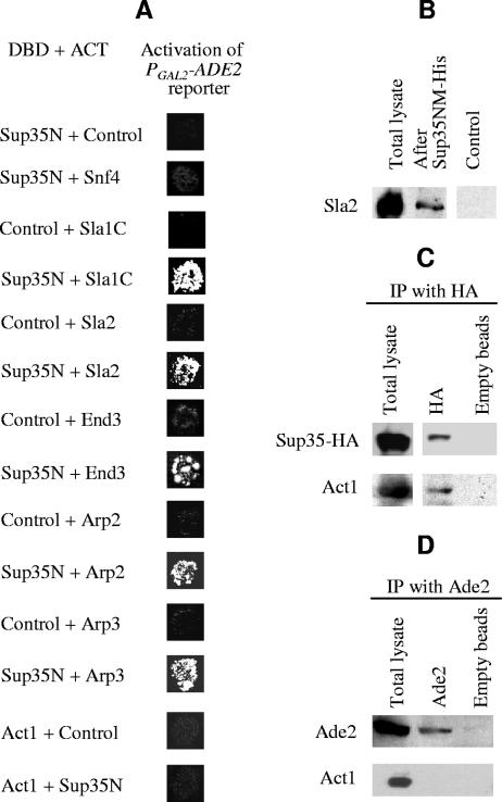
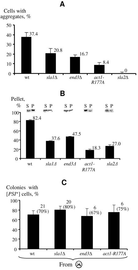
Self-perpetuating protein aggregates transmit prion diseases in mammals and heritable traits in yeast. De novo prion formation can be induced by transient overproduction of the corresponding prion-forming protein or its prion domain. Here, we demonstrate that the yeast prion protein Sup35 interacts with various proteins of the actin cortical cytoskeleton that are involved in endocytosis. Sup35-derived aggregates, generated in the process of prion induction, are associated with the components of the endocytic/vacuolar pathway. Mutational alterations of the cortical actin cytoskeleton decrease aggregation of overproduced Sup35 and de novo prion induction and increase prion-related toxicity in yeast. Deletion of the gene coding for the actin assembly protein Sla2 is lethal in cells containing the prion isoforms of both Sup35 and Rnq1 proteins simultaneously. Our data are consistent with a model in which cytoskeletal structures provide a scaffold for generation of large aggregates, resembling mammalian aggresomes. These aggregates promote prion formation. Moreover, it appears that the actin cytoskeleton also plays a certain role in counteracting the toxicity of the overproduced potentially aggregating proteins.


Figures

FIG. 1.
FIG. 1.
Interactions between Sup35 and cytoskeletal proteins. (A) Two-hybrid assay. DBD, fusion to the DNA binding domain of Gal4; ACT, fusion to the activation domain of Gal4. “Control” refers to plasmids bearing either Gal4DBD or Gal4ACT domains without an insert, respectively. Fusion of the Snf4 protein to the activation domain of Gal4 was used as an additional negative control, showing that Sup35N does not interact with just any randomly chosen yeast protein. Activation of the PGAL-ADE2 reporter construct resulting from a two-hybrid interaction leads to growth on −Ade medium, shown after 7 days of incubation. In all cases, activation of the PGAL-HIS3 reporter construct, resulting in growth on −His medium supplemented with 5 mM aminotriazole, was also tested, with the same result (not shown). All Gal4DBD-Sup35N constructs shown contained Sup35N fused to the N terminus of Gal4DBD; however, in every case, the same result was also observed with a construct containing Sup35N fused to the C terminus of Gal4DBD. In all cases where interaction with Sup35N was detected, the same result was observed with complete Sup35. (B) Sla2 protein was immobilized from yeast extracts on resin containing the His-tagged Sup35NM fragment (Sup35NM-His). Yeast extracts were prepared from the [PSI+ PIN+] yeast strain and run through either Ni2+ resin charged with Sup35NM-His protein (after Sup35NM-His) or control resin charged with the protein prepared from the same Escherichia coli strain bearing the control vector instead of the Sup35NM-His overexpressor (control). Eluates were analyzed by SDS-PAGE and Western blotting, followed by reaction to the Sla2 antibody. A similar result was obtained when the yeast strain was transformed with the plasmid coding for HA-tagged Sla2 and reaction to HA antibody was performed (not shown). (C and D) Actin coprecipitates with Sup35-HA (C) but not with Ade2 (D). Proteins were isolated from the [psi PIN+] yeast strain, expressing the HA-tagged Sup35 from the PCUP1 promoter, and immunoprecipitated with either antibody against HA (recognizing HA-tagged Sup35) (C) or antibody against Ade2 (D). Both total lysates before immunoprecipitation and immunoprecipitates (IP) were run on SDS-PAGE and analyzed by Western blotting, followed by reaction to HA, Ade2, or actin antibody.
FIG. 2.
FIG. 2.
Colocalization of Sup35 aggregates with cytoskeletal components related to the endocytic pathway. (A and B) Different types of aggregates seen after overproduction of Sup35NM-GFP (A) or Sup35-RFP (B) in [psi PIN+] cultures. Cell boundaries are shown. Note that only clumps and dots are seen in the [PSI+] cultures. (C) In the majority of the cases, the presence of aggregates (circle with frowning face) coincides with the ability of cells to form [PSI+] or mosaic [PSI+]/[psi] colonies, while cells with diffuse fluorescence (gray circles) give rise preferentially to [psi] colonies. Cells were isolated by micromanipulation from the [psi PIN+] cultures overexpressing Sup35NM-GFP. Data were combined for the wild-type strain and cytoskeletal mutants, as they were homogenous (see below [Fig. 4C]). Only viable cells are shown (for viability data, see Fig. 5E, below). Bars correspond to standardized errors. (D) Sup35NM-GFP aggregates do not colocalize with cortical actin patches in the [PSI+] strain (upper panel) but do colocalize with some actin patches in the [psi PIN+] strain (middle and bottom panels). (D) “Internal rings” of Sup35NM-GFP (green) are assembled at the periphery of the vacuole in the [psi PIN+] culture. The vacuolar membrane (red) was visualized by staining with the lipophilic dye FM4-64 (see Materials and Methods). Cell boundaries are shown. Images were taken at 45 min after addition of the dye. (F) GFP-tagged Sla1 and Sla2 proteins form patch-like structures in the yeast cells not overexpressing any proteins. (G and H) Sup35-RFP aggregates (red) colocalize with the areas of increased density of Sla1-GFP (G) or Sla2-GFP (H) patches (green) in both [PSI+] and [psi PIN+] cells. In contrast, cells with diffuse distribution of Sup35-RFP do not show colocalization between Sup35-RFP and Sla1 or Sla2 patches. Sup35NM-derived constructs were induced from PCUP1 (A, C, D, and E) or PGAL (B, G, and H), as described in Materials and Methods. Bars, 5 μm in each case.
FIG. 3.
FIG. 3.
[PSI+] induction is decreased in the cytoskeletal mutants. (A and B) Plate assays with PGAL (A) or PCUP1 (B) constructs in the haploid [psi PIN+] strains. Cultures were induced on galactose (A) or copper (B) medium and velveteen replica plated onto −Ade-glucose medium; plate images were taken after 7 days. The sla2Δ strain was not checked in panel A, as it is defective in growth on galactose and some other nonfermentable or poorly fermentable carbon sources. (C) Quantitative assay with PCUP1 constructs. The same cultures as in panel B were induced in the liquid medium and after specified periods of time were plated onto the solid medium. All manipulations were under conditions selective for plasmids. Resulting colonies were scored for [PSI+]. Averages of at least three independent cultures are shown for each strain-plasmid combination, with no less than 70 colonies per culture analyzed per each time point. (D) Levels of Sup35NM-GFP accumulation in the induced cultures are not affected by cytoskeletal mutations. Proteins were isolated after 24 h of copper induction and analyzed by SDS-PAGE and Western blotting, followed by reaction to the Sup35 antibody. Intensities of the bands were determined by densitometry. Levels of Sup35NM-GFP in each culture were normalized by the level of full-size Sup35, produced from a chromosomal copy. Measurements were repeated at least two times with each of at least two different transformants per each strain-plasmid combination. (E) The decrease in [PSI+] induction is not due to decreased detection of [PSI+] colonies. The PGAL-SUP35 induction was performed in the isogenic haploid wild-type and act1-R177A [psi PIN+] strains on solid medium; induced [PSI+] derivatives were detected in a diploid obtained by mating to the wild-type [psi pin] strain, in order to avoid the potential inhibitory effect of act1-R177A on [PSI+] detection, as described in the text. The −Ade plates were photographed after 7 days of incubation. (F) The act1-R177A mutation does not influence efficiency of mating. Equal numbers of cells from the act1-R177A mutant culture and control culture of the isogenic wild-type strain with the integrated HIS3 gene were mixed with a culture of the isogenic wild-type strain of the opposite mating type, used in panel E, and containing plasmid YEp13, which provides the complementary marker LEU2. After 6 h of incubation in YPD medium, serial dilutions were plated onto -His-Leu medium selective for diploids. No difference in diploid frequency was detected between the act1-R177A mutant and wild-type control strain (wt). Standardized errors are shown in panels C and D.
FIG. 4.
FIG. 4.
Sup35 aggregation is decreased in cytoskeletal mutants. (A) Percentage of cells with cytologically detectable Sup35NM-GFP aggregates is decreased in cytoskeletal mutants, compared to the isogenic wild-type strain. Copper induction was for 48 h. Data represent the averages of three independent cultures, with no less than 200 cellsper culture analyzed in each case. (B) Fraction of Sup35NM-GFP pelletable at 9,000 × g is decreased in the cytoskeletal mutants compared to the isogenic wild-type strain. Copper induction was for 24 h. Protein extracts were fractionated into supernatant (S) and pellet (P) by differential centrifugation as described previously (17). Proportion of Sup35NM-GFP in the pellet fraction in each case was determined by densitometry. Averages of at least two repeats with at least two different transformants per each strain are shown. (C) Viable cells with detectable fluorescent Sup35NM-GFP aggregates (circle with frowning face), isolated by micromanipulation from the wild-type and mutant cultures, gave rise to colonies containing [PSI+] cells with similar frequencies. For viability data, see below (Fig. 5E). Standardized errors are shown.
FIG. 5.
FIG. 5.
Prion-related toxicity of excess Sup35 is increased in the cytoskeletal mutants. (A) Toxicity of excess Sup35 (↑Sup35), overexpressed from the PGAL promoter, is increased in [PSI+] strains bearing the cytoskeletal mutations. Expression of the Sup35C region lacking the prion domain (↑Sup35C) from the PCUP1 promoter simultaneously with complete Sup35 ameliorates toxicity in the wild-type strain but not in the mutant strains. “Control” refers to the isogenic strains not overexpressing Sup35 or Sup35C. Plates were photographed after 4 days of incubation. (B and C) Toxicity of Sup35NM-GFP, overproduced from PCUP1, is increased in [PSI+] strains (B) and [psi PIN+] strains (C) with cytoskeletal mutations. At each time point, concentrations of CFU in each culture growing in the liquid medium with copper were determined by plating onto the solid medium without additional copper. To account for the potential general growth defect of cytoskeletal mutants that is independent of Sup35NM-GFP accumulation, every CFU concentration of a Sup35NM-GFP-overproducing culture was divided by the CFU concentration of the isogenic culture with empty vector without SUP35NM-GFP, grown under the same conditions. All manipulations were performed under conditions selective for plasmids. (D) Toxicity of high levels of overproduced Sup35 is increased in [psi PIN+] strains with cytoskeletal mutations. The multicopy plasmid pEMBL-yex-SUP35, bearing the SUP35 gene under its own promoter (↑↑Sup35) was amplified on −Leu medium, selecting for the partially defective LEU2-d allele, as described in the text. Plates were photographed after 4 days of incubation. No observable differences in growth were detected for isogenic transformants bearing the control LEU2-d plasmid pEMBL-yex without the SUP35 gene (not shown). In both panels A and D, at least eight independent transformants were analyzed per each strain-plasmid combination, with the same result. Therefore, differences in growth observed between wild-type and any mutant strain were statistically significant (P < 0.01). (E) Viability of cells with Sup35NM-GFP aggregates (circle with frowning face) and diffuse fluorescence (gray circle) in the wild-type strain and cytoskeletal mutants. Cells were sorted by micromanipulation from the [psi PIN+] cultures overproducing Sup35NM-GFP and incubated for 2 days on YPD medium. At least 20 cells of each type were analyzed per each culture. Standardized errors are shown in panels B, C, and E. In each case where error bars cannot be seen, the error was less than the size of the symbol.
FIG. 6.
FIG. 6.
Model for the effects of the actin cytoskeleton and endocytic pathway on Sup35 aggregation and prion formation. See comments in the text.

References

    1. Allen, K. D., R. D. Wegrzyn, T. A. Chernova, S. Müller, G. P. Newnam, P. A. Winslett, K. B. Wittich, K. D. Wilkinson, and Y. O. Chernoff. 2005. Hsp70 chaperones as modulators of prion life cycle: novel effects of Ssa and Ssb on the Saccharomyces cerevisiae prion [PSI+]. Genetics 169:1227-1242. - PMC - PubMed
    1. Ayscough, K. R. 2000. Endocytosis and the development of cell polarity in yeast require a dynamic F-actin cytoskeleton. Curr. Biol. 10:1587-1590. - PubMed
    1. Ayscough, K. R., J. J. Eby, T. Lila, H. Dewar, K. G. Kozminski, and D. G. Drubin. 1999. Sla1p is a functionally modular component of the yeast cortical actin cytoskeleton required for correct localization of both Rho1p-GTPase and Sla2p, a protein with talin homology. Mol. Biol. Cell 10:1061-1075. - PMC - PubMed
    1. Bailleul, P. A., G. P. Newnam, J. N. Steenbergen, and Y. O. Chernoff. 1999. Genetic study of interactions between the cytoskeletal assembly protein sla1 and prion-forming domain of the release factor Sup35 (eRF3) in Saccharomyces cerevisiae. Genetics 153:81-94. - PMC - PubMed
    1. Bailleul-Winslett, P. A., G. P. Newnam, R. D. Wegrzyn, and Y. O. Chernoff. 2000. An antiprion effect of the anticytoskeletal drug latrunculin A in yeast. Gene Expr. 9:145-156. - PMC - PubMed

Publication types

LinkOut - more resources