Pannus resolution after occipitocervical fusion in a non-rheumatoid atlanto-axial instability
- PMID: 16382308
- PMCID: PMC3489286
- DOI: 10.1007/s00586-005-0969-4
Pannus resolution after occipitocervical fusion in a non-rheumatoid atlanto-axial instability
Abstract
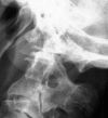
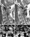
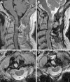
Periodontoid pseudotumor or pannus is considered to be an inflammatory mass most frequently associated with rheumatoid arthritis. Transoral resection of the pannus has been the treatment of choice for patients with associated myelopathy, followed in many instances by posterior stabilization. However, some authors have reported resolution of pannus associated with rheumatoid arthritis and other forms of chronic atlanto-axial instability only after posterior stabilization. We report a case of a 69-year-old man who presented with a rapidly progressing myelopathy due to a retro-odontoid mass produced by chronic atlanto-axial instability associated with an occipital assimilation of C1 and tight posterior fossa. An urgent posterior fossa craniectomy followed by occipitocervical fixation was performed. After surgery, the patient's clinical condition improved and 1 year after surgery was asymptomatic, walked without any help and had normal strength. Control MR showed complete resolution of the retro-odontoid pannus.
Figures


Similar articles
-
Clinical and Radiographic Outcomes of Atlantoaxial or Occipitocervical Fixation and Fusion in Patients With Cervical Myelopathy due to Idiopathic Retro-Odontoid Pseudotumor.Oper Neurosurg. 2025 Mar 5;29(1):102-108. doi: 10.1227/ons.0000000000001512. Oper Neurosurg. 2025. PMID: 40042300
-
Disappearance of degenerative, non-inflammatory, retro-odontoid pseudotumor following posterior C1-C2 fixation: case series and review of the literature.Eur Spine J. 2013 Nov;22 Suppl 6(Suppl 6):S879-88. doi: 10.1007/s00586-013-3004-1. Epub 2013 Sep 19. Eur Spine J. 2013. PMID: 24048650 Free PMC article.
-
Pannus regression after posterior decompression and occipito-cervical fixation in occipito-atlanto-axial instability due to rheumatoid arthritis: case report and literature review.Clin Neurol Neurosurg. 2013 Feb;115(2):111-6. doi: 10.1016/j.clineuro.2012.04.018. Epub 2012 May 19. Clin Neurol Neurosurg. 2013. PMID: 22609344 Review.
-
Reduction of rheumatoid periodontoid pannus following posterior occipito-cervical fusion visualised by magnetic resonance imaging.Br J Neurosurg. 1988;2(3):315-20. doi: 10.3109/02688698809001001. Br J Neurosurg. 1988. PMID: 3267314
-
Retro-odontoid Degenerative Pseudotumour Causing Spinal Cord Compression and Myelopathy: Current Evidence on the Role of Posterior C1-C2 Fixation in Treatment.Acta Neurochir Suppl. 2019;125:259-264. doi: 10.1007/978-3-319-62515-7_37. Acta Neurochir Suppl. 2019. PMID: 30610331 Review.
Cited by
-
Regression of Disc-Osteophyte Complexes Following Laminoplasty Versus Laminectomy with Fusion for Cervical Spondylotic Myelopathy.Int J Spine Surg. 2017 Jun 12;11(3):17. doi: 10.14444/4017. eCollection 2017. Int J Spine Surg. 2017. PMID: 28765801 Free PMC article.
-
Exploration for reliable radiographic assessment method for hinge-like hypermobility at atlanto-occipital joint.Eur Spine J. 2018 Jun;27(6):1303-1308. doi: 10.1007/s00586-017-5349-3. Epub 2017 Oct 20. Eur Spine J. 2018. PMID: 29052813
-
Retro-Odontoid Pseudotumor with Cervical Medullary Compression: A Case Report.Spartan Med Res J. 2018 Apr 27;3(1):6768. doi: 10.51894/001c.6768. Spartan Med Res J. 2018. PMID: 33655134 Free PMC article.
-
Retroodontoid Pseudotumor Related to Development of Myelopathy Secondary to Atlantoaxial Instability on Os Odontoideum.Case Rep Radiol. 2018 Sep 30;2018:1658129. doi: 10.1155/2018/1658129. eCollection 2018. Case Rep Radiol. 2018. PMID: 30363967 Free PMC article.
-
Thinking beyond pannus: a review of retro-odontoid pseudotumor due to rheumatoid and non-rheumatoid etiologies.Skeletal Radiol. 2019 Oct;48(10):1511-1523. doi: 10.1007/s00256-019-03187-z. Epub 2019 Mar 13. Skeletal Radiol. 2019. PMID: 30868232 Review.
References
-
- Crockard HA, Pozo JL, Ransford AO, Stevens JM, Kendall BE, Essigman WK. Transoral decompression and posterior fusion for rheumatoid atlanto-axial subluxation. J Bone Joint Surg Br. 1986;68:350–356. - PubMed
-
- Joly-Torta MC, Martín-Ferrer S, Rimbau-Muñoz J, Domínguez C. Reducción de las masas periodontoideas tras artrodesis posterior: revisión a propósito de 2 casos no vinculados a artritis reumatoide. Neurocirugía (Astur) 2004;15:553–564. - PubMed
Publication types
MeSH terms
LinkOut - more resources
Full Text Sources
Medical

