Review
. 2006:(95):193-211.
doi: 10.1007/3-7643-7417-9_15.

Plasticity of neuropeptide Y in the dentate gyrus after seizures, and its relevance to seizure-induced neurogenesis

Affiliations
Review

Plasticity of neuropeptide Y in the dentate gyrus after seizures, and its relevance to seizure-induced neurogenesis

Helen E Scharfman et al. EXS. 2006.

Abstract

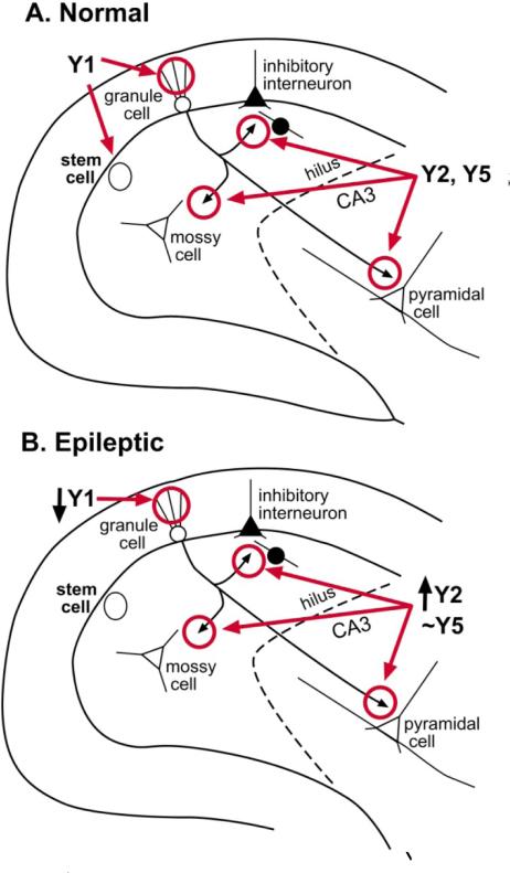
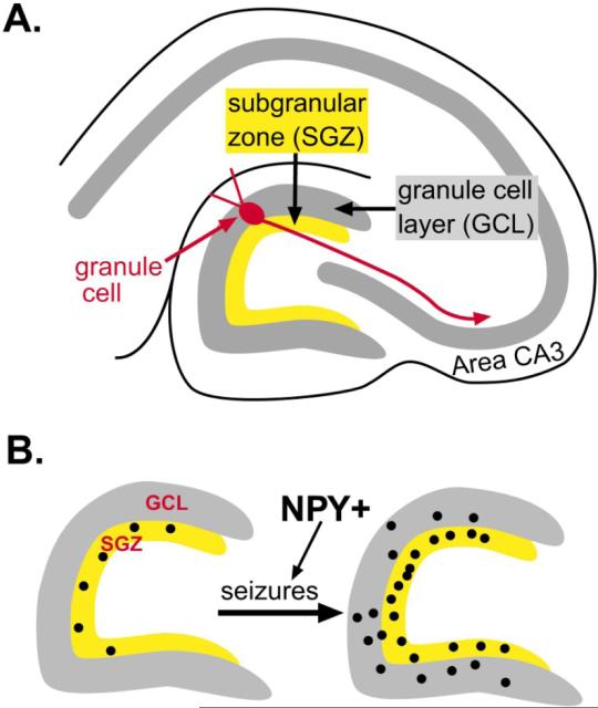
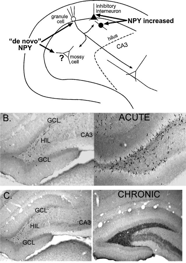
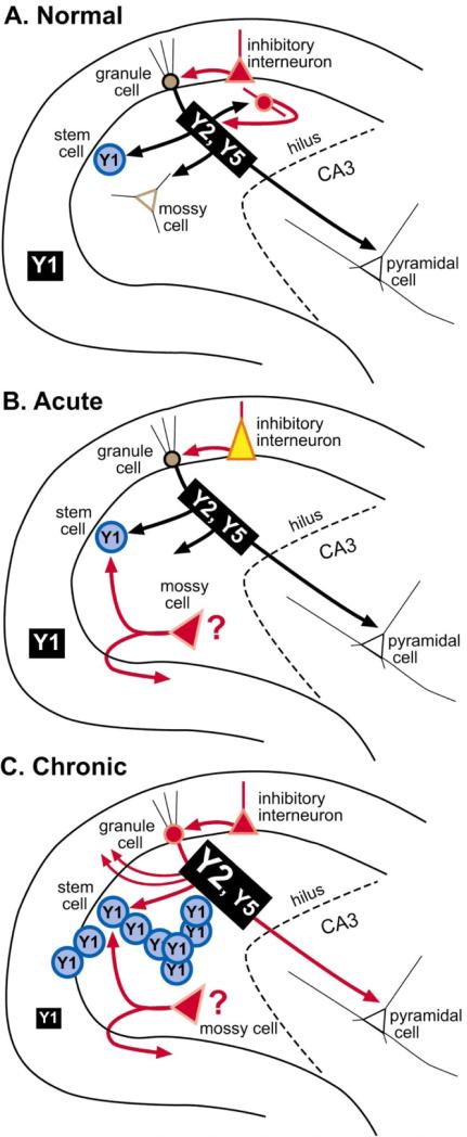
In summary, NPY is clearly an important peptide in the adult rat dentate gyrus because it has the potential to influence synaptic transmission and neurogenesis. It may even have other functions, as yet undiscovered, mediated by glia or vasculature. The remarkable plasticity of NPY puts it in a position to allow dentate gyrus function to be modified in a changing environment. The importance of this plasticity in the context of epilepsy cannot be emphasized enough. It could help explain a range of observations about epilepsy that currently is poorly understood. For example, rapid increases in NPY could mediate postictal depression, the period of depression that can last for several hours after generalized seizures. It may mediate the "priming effect," which is a reduction in seizure threshold following an initial period of seizures. Finally, it could contribute to the resistance of dentate granule cells to degeneration after seizures. However, despite the focus in this review on seizure-induced changes, the changes described here also appear to occur after other types of manipulations, which considerably broadens the scope of NPY's role in the brain.


Figures

Figure 1
Figure 1
Schematic of the adult rat dentate gyrus. A transverse section through the rat dentate gyrus in the horizontal plane illustrates its location in the hippocampus. The inset diagrams the locations of the different cell types and lamination of the dentate gyrus.
Figure 2
Figure 2
Normal NPY expression in the adult dentate gyrus. TOP: A schematic shows the normal distribution of NPY protein is in various inhibitory neurons. BOTTOM: A micrograph showing expression of NPY in a normal male adult rat. The neurons that are NPY-immunoreactive have somata mainly in the granule cell layer (GCL) and hilus (HIL), and co-express GABA. DG = dentate gyrus. MOL = molecular layer.
Figure 3
Figure 3
The distribution of NPY receptors in the normal and epileptic rat dentate gyrus. NPY receptor distribution of a normal adult rat (A) and an adult rat that has had severe continuous seizures (status epilepticus; B) are shown. The distributions appears similar regardless of the methods to induce status, and seizures last for 1 or more hours. Methods that are commonly used to induce status include the convulsant pilocarpine or kainic acid, or electrical stimulation. After status epilepticus, spontaneous repetitive seizures develop within weeks, and persist for the life of the animal (i.e., epileptogenesis occurs). For further explanation and references, see text.
Figure 4
Figure 4
Seizure-induced neurogenesis in the rat dentate gyrus. A. A schematic illustrates the area of the dentate gyrus where progenitors are located in the adult rat, the subgranular zone. B. A schematic illustrates the increase in progenitors, labeled by the mitotic marker bromodeoxyuridine (BrdU), after seizures. As discussed in the text, NPY appears to facilitate this process.
Figure 5
Figure 5
Changes in NPY expression after seizures. A. A diagram is used to illustrate where NPY protein increases in the adult rat dentate gyrus after seizures. It appears to increase in inhibitory neurons, and also develop in neurons such as granule cells and mossy cells that do not normally express the protein (de novo expression). B. The increase in NPY protein in the dentate gyrus is shown using an antibody to NPY. Left: Saline control. Right: 1 day after status epilepticus. C. Increased NPY in mossy fibers is illustrated after chronic seizures. Left: Saline control. Right: 2 months after status epilepticus. Spontaneous seizures were observed in the animal that had status epilepticus.
Figure 6
Figure 6
De novo expression of NPY in hilar cells after seizures. NPY immunoreactivity in the den-tate gyrus of a pilocarpine-treated rat that had no behavioral seizures (A-C) compared to a pilocarpine-treated rat that had I h of status epilepticus and subsequently had spontaneous seizures (D-F). Both animals were killed 2 months after pilocarpine treatment. A-C. The normal pattern of NPY immunoreactivity includes NPY expression in many hilar cells and fibers, as well as fibers in the outer molecular layer (A). B and C show NPY-immunoreactive hilar cells at higher magnification. GCL = granule cell layer; HIL = hilus. D-F. NPY immunoreactivity in the epileptic rat shows increased immunoreactivity in hilar cells and in fibers (D; same magnification as A). In addition, a novel band of staining in the inner molecular layer is present, a reflection of mossy fiber sprouting. E and F show higher magnification of hilar cells that are NPY-immunoreactive. Note the large size of these cells and irregular, large primary dendrites (arrows) relative to the normal NPY-immunoreactive cells shown in E and F (magnification is the same in B, C, E, F). It is assumed from these changes in immunoreactivity that the cells in the normal hilus die due to seizure-induced neuronal death, and the residual surviving cells, including some mossy cells, develop NPY immunoreactivity.
Figure 7
Figure 7
Summary of NPY plasticity in the rat dentate gyrus after seizures. A summary of the changes that occur in protein and receptor expression after acute seizures and further changes after chronic seizures. A. Normal condition B. After acute seizures, NPY protein increases in inhibitory neurons, although some may also be lost due to seizure-induced neuronal death, and some may sprout collaterals, making the new NPY intemeurons network potentially novel. In addition, some hilar cells that do not normally express NPY protein may begin to do so, such as surviving mossy cells. C. After chronic seizures, granule cells and their axons, the mossy fibers, express NPY and sprout into the inner molecular layer. Y2, and possibly Y5 receptors, increase in mossy fibers, and Y1 receptors in the molecular layer appear to decrease. In addition, seizures increase the proliferation of granule cells from progenitors in the subgranular zone which express the Y1 receptor; this is likely to occur during a window between day 3–4 and 30 after status epilepticus, at least in the case of pilocarpine-induced status epilepticus [32, 34]. This coincides with the period when spontaneous seizures are beginning to occur, suggesting a role for seizure-induced neurogenesis in epileptogenesis [33].

References

    1. Abe H, Watanabe M, Yamakuni T, Kuwano R, Takahashi Y, Kondo H. Localization of gene expression of calbindin in the brain of adult rats. Neurosci Lett. 1992;138:211–215. - PubMed
    1. Frederickson CJ, Danscher G. Zinc-containing neurons in hippocampus and related CNS structures. Prog Brain Res. 1990;83:71–84. - PubMed
    1. Gall C, Brecha N, Karten HJ, Chang KJ. Localization ofenkephalin-like immunoreactivity to identified axonal and neuronal populations of the rat hippocampus. J Comp Neurol. 1981;198:335–350. - PubMed
    1. Freund TF, Hajos N, Acsady L, Gores TJ, Katona I. Mossy cells of the rat dentate gyrus are immunoreactive for calcitonin gene-related peptide (CGRP). Eur J Neurosci. 1997;9:1815–1830. - PubMed
    1. Freund TF, Bazsaki G. Intemeurons of the hippocampus. Hippocampus. 1996;6:347–470. - PubMed

Publication types