. 2006 Jan 10;103(2):305-10.
doi: 10.1073/pnas.0507109103. Epub 2005 Dec 29.

Mechanism of bacterial cell-surface attachment revealed by the structure of cellulosomal type II cohesin-dockerin complex

Affiliations

Mechanism of bacterial cell-surface attachment revealed by the structure of cellulosomal type II cohesin-dockerin complex

Jarrett J Adams et al. Proc Natl Acad Sci U S A. .

Abstract

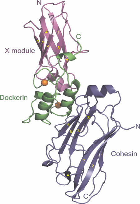
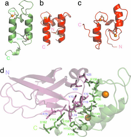
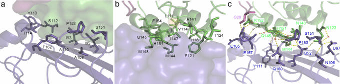
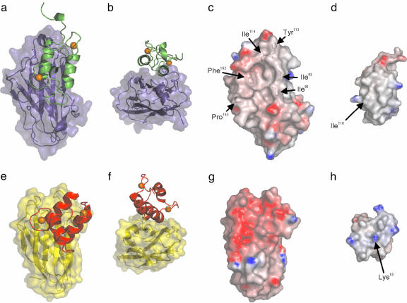
Bacterial cell-surface attachment of macromolecular complexes maintains the microorganism in close proximity to extracellular substrates and allows for optimal uptake of hydrolytic byproducts. The cellulosome is a large multienzyme complex used by many anaerobic bacteria for the efficient degradation of plant cell-wall polysaccharides. The mechanism of cellulosome retention to the bacterial cell surface involves a calcium-mediated protein-protein interaction between the dockerin (Doc) module from the cellulosomal scaffold and a cohesin (Coh) module of cell-surface proteins located within the proteoglycan layer. Here, we report the structure of an ultra-high-affinity (K(a) = 1.44 x 10(10) M(-1)) complex between type II Doc, together with its neighboring X module from the cellulosome scaffold of Clostridium thermocellum, and a type II Coh module associated with the bacterial cell surface. Identification of X module-Doc and X module-Coh contacts reveal roles for the X module in Doc stability and enhanced Coh recognition. This extremely tight interaction involves one face of the Coh and both helices of the Doc and comprises significant hydrophobic character and a complementary extensive hydrogen-bond network. This structure represents a unique mechanism for cell-surface attachment in anaerobic bacteria and provides a rationale for discriminating between type I and type II Coh modules.


Figures

Fig. 1.
Fig. 1.
Structure of the type II Coh-XDoc complex. Ribbon representation of the complex with the type II Coh module in blue, type II Doc in green, and X module in magenta. The β-strands of the X module and type II Coh are numbered in yellow. The N and C termini are labeled accordingly, and the Ca2+ ions are depicted as orange spheres.
Fig. 2.
Fig. 2.
Doc conformations and XDoc interface. The structures of type II Doc from the type II Coh-XDoc complex (a); type I Doc from the type I Coh-Doc complex (21) (b); and isolated type I Doc (15) (c) illustrate the differences in loop conformations. Coordinating residues at position 1 and 12 of each Ca2+-binding loop as stick models, with the Asp at position 12 in each loop of the type II-Doc structure labeled with one-letter code and the corresponding residue number. (d) The XDoc illustration shows the module-module contacts. Interface residues from the X module and type II Doc are depicted as magenta and green stick models, respectively, on the backbone ribbon representation of the XDoc structure.
Fig. 3.
Fig. 3.
Type II Coh-XDoc complex interface contacts. (a) Ribbon representation of type II Coh, displaying hydrophobic interface residues as stick models on the molecular-surface representation of XDoc. (b) Ribbon representation of XDoc, displaying hydrophobic interface residues as stick models on the molecular-surface representation of type II Coh. (c) Interface hydrogen-bond network, with water molecules shown as red X and hydrogen-bond contacts as yellow dashed lines. Type II Coh, Doc, and X module are colored blue, green, and magenta, respectively. Residues depicted as stick models are labeled accordingly.
Fig. 4.
Fig. 4.
Interaction surfaces of type I- and type II Coh-Doc complexes. Ribbon representations of type II Doc (green) (a) on the molecular surface of the type II Coh (blue) and type I Doc (red) (e) on the molecular surface of type I Coh (yellow) (21). Representations in b and f have been rotated clockwise 90° around the x axis, followed by a 180° clockwise rotation around the z axis. Electrostatic surface potential representations of C. thermocellum type II Coh (c), C. thermocellum type II Doc (d), C. thermocellum type I Coh (21) (g), and C. thermocellum type I Doc (21) (h). Positive regions are shown in blue and negative regions in red. Residues contributing to the hydrophobic surface character of C. thermocellum type II Coh are labeled accordingly. The location of Ile-118 on the surface of the type II Doc and the analogous residue in the type I Doc (Lys-18) are identified. The electrostatic surface potentials were calculated in grasp (47) and are contoured from -14 (red) to +14 (blue). Ca2+ ions are shown as orange spheres.

References

    1. Bayer, E. A., Belaich, J. P., Shoham, Y. & Lamed, R. (2004) Annu. Rev. Microbiol. 58, 521-554. - PubMed
    1. Bayer, E. A., Chanzy, H., Lamed, R. & Shoham, Y. (1998) Curr. Opin. Struct. Biol. 8, 548-557. - PubMed
    1. Beguin, P. & Lemaire, M. (1996) Crit. Rev. Biochem. Mol. Biol. 31, 201-236. - PubMed
    1. Demain, A. L., Newcomb, M. & Wu, J. H. (2005) Microbiol. Mol. Biol. Rev. 69, 124-154. - PMC - PubMed
    1. Doi, R. H. & Kosugi, A. (2004) Nat. Rev. Microbiol. 2, 541-551. - PubMed

Publication types

MeSH terms

Substances

Associated data

LinkOut - more resources