Spectrum and frequency of mutations in IMPDH1 associated with autosomal dominant retinitis pigmentosa and leber congenital amaurosis
- PMID: 16384941
- PMCID: PMC2581444
- DOI: 10.1167/iovs.05-0868
Spectrum and frequency of mutations in IMPDH1 associated with autosomal dominant retinitis pigmentosa and leber congenital amaurosis
Abstract
Purpose: The purpose of this study was to determine the frequency and spectrum of inosine monophosphate dehydrogenase type I (IMPDH1) mutations associated with autosomal dominant retinitis pigmentosa (RP), to determine whether mutations in IMPDH1 cause other forms of inherited retinal degeneration, and to analyze IMPDH1 mutations for alterations in enzyme activity and nucleic acid binding.
Methods: The coding sequence and flanking intron/exon junctions of IMPDH1 were analyzed in 203 patients with autosomal dominant RP (adRP), 55 patients with autosomal recessive RP (arRP), 7 patients with isolated RP, 17 patients with macular degeneration (MD), and 24 patients with Leber congenital amaurosis (LCA). DNA samples were tested for mutations by sequencing only or by a combination of single-stranded conformational analysis and by sequencing. Production of fluorescent reduced nicotinamide adenine dinucleotide (NADH) was used to measure enzymatic activity of mutant IMPDH1 proteins. The affinity and the specificity of mutant IMPDH1 proteins for single-stranded nucleic acids were determined by filter-binding assays.
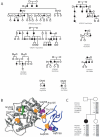
Results: Five different IMPDH1 variants, Thr116Met, Asp226Asn, Val268Ile, Gly324Asp, and His 372Pro, were identified in eight autosomal dominant RP families. Two additional IMPDH1 variants, Arg105Trp and Asn198Lys, were found in two patients with isolated LCA. None of the novel IMPDH1 mutants identified in this study altered the enzymatic activity of the corresponding proteins. In contrast, the affinity and/or the specificity of single-stranded nucleic acid binding were altered for each IMPDH1 mutant except the Gly324Asp variant.
Conclusions: Mutations in IMPDH1 account for approximately 2% of families with adRP, and de novo IMPDH1 mutations are also rare causes of isolated LCA. This analysis of the novel IMPDH1 mutants substantiates previous reports that IMPDH1 mutations do not alter enzyme activity and demonstrates that these mutants alter the recently identified single-stranded nucleic acid binding property of IMPDH. Studies are needed to further characterize the functional significance of IMPDH1 nucleic acid binding and its potential relationship to retinal degeneration.
Figures


References
-
- Kennan A, Aherne A, Palfi A, et al. Identification of an IMPDH1 mutation in autosomal dominant retinitis pigmentosa (RP10) revealed following comparative microarray analysis of transcripts derived from retinas of wild-type and Rho(-/-) mice. Hum Mol Genet. 2002;11:547–557. - PubMed
-
- Gu JJ, Spychala J, Mitchell BS. Regulation of the human inosine monophosphate dehydrogenase type I gene. Utilization of alternative promoters. J Biol Chem. 1997;272:4458–4466. - PubMed
-
- Senda M, Natsumeda Y. Tissue-differential expression of two distinct genes for human IMP dehydrogenase (E.C. 1.1.1.205) Life Sci. 1994;54:1917–1926. - PubMed
Publication types
MeSH terms
Substances
Grants and funding
LinkOut - more resources
Full Text Sources
Other Literature Sources
Molecular Biology Databases
Research Materials

