. 2006 Jan;188(2):370-7.
doi: 10.1128/JB.188.2.370-377.2006.

Nanoscale characterization and determination of adhesion forces of Pseudomonas aeruginosa pili by using atomic force microscopy

Affiliations

Nanoscale characterization and determination of adhesion forces of Pseudomonas aeruginosa pili by using atomic force microscopy

Ahmed Touhami et al. J Bacteriol. 2006 Jan.

Abstract

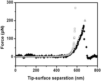
Type IV pili play an important role in bacterial adhesion, motility, and biofilm formation. Here we present high-resolution atomic force microscopy (AFM) images of type IV pili from Pseudomonas aeruginosa bacteria. An individual pilus ranges in length from 0.5 to 7 microm and has a diameter from 4 to 6 nm, although often, pili bundles in which the individual filaments differed in both length and diameter were seen. By attaching bacteria to AFM tips, it was possible to fasten the bacteria to mica surfaces by pili tethers. Force spectra of tethered pili gave rupture forces of 95 pN. The slopes of force curves close to the rupture force were nearly linear but showed little variation with pilus length. Furthermore, force curves could not be fitted with wormlike-chain polymer stretch models when using realistic persistence lengths for pili. The observation that the slopes near rupture did not depend on the pili length suggests that they do not represent elastic properties of the pili. It is possible that this region of the force curves is determined by an elastic element that is part of the bacterial wall, although further experiments are needed to confirm this.


Figures

FIG. 1.
FIG. 1.
(A) EM image of an AFM tip coated with poly-l-lysine (bar, 840 nm). (B) EM image of an AFM tip decorated with bacteria near the base of the tip. Bacteria generally attached on the sides of the pyramidal tip or more frequently near the base (bar, 640 nm).
FIG. 2.
FIG. 2.
TEM images of pili for P. aeruginosa PAO1. (A) Individual entangled pili. Sharp bends in the pili (arrow) were produced through the sample drying process. (B) Example of the tendency for pili to form bundles.
FIG. 3.
FIG. 3.
(A) AFM image of the pole region of a P. aeruginosa PAO1 bacterium showing a flagellum and several straight pili. (B) Pili embedded in the layer of surface deposit. In some regions (black arrow), pili appear to be nearly submerged in a smooth region of the deposit. The white arrow points to a pili bundle. (C) Example of a bare region of the mica substrate traversed by pili fibers. (D) Cross section of the pilus in panel C along the line indicated. Such cross sections had pili diameters of 4 to 6 nm. Scale bars, 250 nm.
FIG. 4.
FIG. 4.
(A) AFM image of a single straight pilus. The inset represents an enlarged region of the pilus and shows a helical fine structure on the pilus surface. (B) A frequently observed branching of pili. Scale bar, 300 nm.
FIG. 5.
FIG. 5.
(A) Collection of force spectra (retraction) taken with bacterium-coated tips over mica surfaces. Tip retraction speeds were 3 μm/s, and the spectra from a to f were arranged in order of increasing retraction distance needed to apply tension on the pili.
FIG. 6.
FIG. 6.
(A) Distribution of observed rupture forces for pili force spectra over mica surfaces. A mean rupture force of 95 pN was obtained. (B) Distribution of corresponding piezo retraction distances needed for rupture. Corresponding pili lengths depend on the (unknown) positions of the bacteria on the AFM cantilever. (C) Distribution of piezo retraction distances for bond rupture for poly-l-lysine-coated AFM tip. No rupture lengths in excess of 600 nm were observed. The inset shows a typical force spectrum for poly-l-lysine. (D) Distribution of piezo retraction distances for bond rupture for strain PAK-Δpil without pili. No rupture lengths in excess of 100 nm were observed. The inset shows a typical force spectrum for strain PAK-Δpil.
FIG. 7.
FIG. 7.
Drawing showing possible ways in which pili can tether bacteria to a surface. In panel A, the tip touches the surface and long pili from bacteria near the tip apex or near the tip base are folded and adhere with their distal end to the mica surface. (B) On tip retraction, pili straighten out, and the shortest pili exerts a force on the cantilever, thus producing a force signature in the force spectrum. (C) On further retraction, some part of the bacterium-pili-substrate complex breaks (most likely the pili-substrate bond) and the cantilever returns to its neutral position. On further retraction, the above process will be repeated with the next longest pili. (D) An alternate model, discussed in the text, of the form of the tether that anchors bacteria to the substrate. Here, several turns of short pili fibers are unraveled and the constituent pilin molecules are denatured, thus forming a long amino acid chain with much lower persistence length than whole pili.
FIG. 8.
FIG. 8.
Sections of force curves a, b, c, d, e, and f in Fig. 5 superimposed to illustrate the similarity of the slopes of the nearly linear regions near rupture. Individual traces were shifted horizontally until curves near the principal adhesion peaks overlapped at a force of 75 pN. The extra hump at 920 nm (derived from trace d in Fig. 5) is most likely caused by adhesion of two pili of nearly the same length.
FIG. 9.
FIG. 9.
⧫, cantilever force as function of tip-surface separation distance for the first adhesion feature in trace a in Fig. 5. The zero for the horizontal axis was taken as the first position in the traces in Fig. 5 where the cantilever deflection on retraction was zero. □, WLC model with persistence length of 0.8 nm and contour length of 600 nm. For larger values for L, the curvature of the force curve as it approaches the nearly linear region could not be reproduced for reasonable persistence lengths. Δ, WLC model with a p of 0.8 nm, a L of 600 nm, and a linear spring with spring constant of 0.002 N/m. With the same linear spring included, the WLC model could be fitted to the main features in the other force curves shown in Fig. 5.

References

    1. Bieber, D., S. W. Ramer, C. Y. Wu, W. J. Murray, T. Tobe, R. Fernandez, and G. K. Schoolnik. 1998. Type IV pili, transient bacterial aggregates, and virulence of enteropathogenic Escherichia coli. Science 280:2114-2118. - PubMed
    1. Boyd, J., T. Koga, and S. Lory. 1994. Identification and characterization of PilS, an essential regulator of pilin expression in pseudomonas aeruginosa. Mol. Gen. Genet. 243:565-574. - PubMed
    1. Craig, L., M. E. Pique, and J. A. Tainer. 2004. Type IV pilus structure and bacterial pathogenicity. Nat. Rev. Microbiol. 2:363-378. - PubMed
    1. Craig, L., R. K. Taylor, M. E. Pique, B. D. Adair, A. S. Arvai, M. Singh, S. J. Lloyed, D. S. Shin, E. D. Getzoff, M. Yeager, K. T. Forest, and J. A. Tainer. 2003. Type IV pilin structure and assembly: X-ray and EM analyses of Vibrio cholaere toxin-coregulated pilus and Pseudomonas aeruginosa PAK pilin. Mol. Cell 11:1139-1150. - PubMed
    1. Dufrêne, Y. F. 2004. Using nanotechniques to explore microbial surfaces. Nat. Rev. Microbiol. 2:451-460. - PubMed

Publication types

MeSH terms

LinkOut - more resources