

Whole-genome plasticity among Mycobacterium avium subspecies: insights from comparative genomic hybridizations
- PMID: 16385061
- PMCID: PMC1347307
- DOI: 10.1128/JB.188.2.711-723.2006
Whole-genome plasticity among Mycobacterium avium subspecies: insights from comparative genomic hybridizations
Abstract
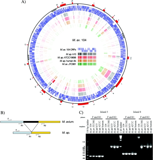
Infection with Mycobacterium avium subsp. paratuberculosis causes Johne's disease in cattle and is also implicated in cases of Crohn's disease in humans. Another closely related strain, M. avium subsp. avium, is a health problem for immunocompromised patients. To understand the molecular pathogenesis of M. avium subspecies, we analyzed the genome contents of isolates collected from humans and domesticated or wildlife animals. Comparative genomic hybridizations indicated distinct lineages for each subspecies where the closest genomic relatedness existed between M. avium subsp. paratuberculosis isolates collected from human and clinical cow samples. Genomic islands (n = 24) comprising 846 kb were present in the reference M. avium subsp. avium strain but absent from 95% of M. avium subsp. paratuberculosis isolates. Additional analysis identified a group of 18 M. avium subsp. paratuberculosis-associated islands comprising 240 kb that were absent from most of the M. avium subsp. avium isolates. Sequence analysis of DNA regions flanking the genomic islands identified three large inversions in addition to several small inversions that could play a role in regulation of gene expression. Analysis of genes encoded in the genomic islands reveals factors that are probably important for various mechanisms of virulence. Overall, M. avium subsp. avium isolates displayed a higher level of genomic diversity than M. avium subsp. paratuberculosis isolates. Among M. avium subsp. paratuberculosis isolates, those from wildlife animals displayed the highest level of genomic rearrangements that were not observed in other isolates. The presented findings will affect the future design of diagnostics and vaccines for Johne's and Crohn's diseases and provide a model for genomic analysis of closely related bacteria.
Figures


Similar articles
-
Genomic homogeneity between Mycobacterium avium subsp. avium and Mycobacterium avium subsp. paratuberculosis belies their divergent growth rates.BMC Microbiol. 2003 May 9;3:10. doi: 10.1186/1471-2180-3-10. BMC Microbiol. 2003. PMID: 12740027 Free PMC article.
-
Comparative transcriptional analysis of human macrophages exposed to animal and human isolates of Mycobacterium avium subspecies paratuberculosis with diverse genotypes.Infect Immun. 2006 Nov;74(11):6046-56. doi: 10.1128/IAI.00326-06. Infect Immun. 2006. PMID: 17057086 Free PMC article.
-
Genome scale comparison of Mycobacterium avium subsp. paratuberculosis with Mycobacterium avium subsp. avium reveals potential diagnostic sequences.J Clin Microbiol. 2002 Apr;40(4):1303-10. doi: 10.1128/JCM.40.4.1303-1310.2002. J Clin Microbiol. 2002. PMID: 11923349 Free PMC article.
-
[Mycobacterium avium subsp. paratuberculosis in food and its relationship with Crohn's disease].Rev Argent Microbiol. 2007 Jan-Mar;39(1):57-68. Rev Argent Microbiol. 2007. PMID: 17585661 Review. Spanish.
-
Epidemiological evidence for Mycobacterium avium subspecies paratuberculosis as a cause of Crohn's disease.Epidemiol Infect. 2007 Oct;135(7):1057-68. doi: 10.1017/S0950268807008448. Epub 2007 Apr 20. Epidemiol Infect. 2007. PMID: 17445316 Free PMC article. Review.
Cited by
-
Superior Protection from Live-Attenuated Vaccines Directed against Johne's Disease.Clin Vaccine Immunol. 2017 Jan 5;24(1):e00478-16. doi: 10.1128/CVI.00478-16. Print 2017 Jan. Clin Vaccine Immunol. 2017. PMID: 27806993 Free PMC article.
-
Direct PCR on Tissue Samples To Detect Mycobacterium tuberculosis Complex: an Alternative to the Bacteriological Culture.J Clin Microbiol. 2021 Jan 21;59(2):e01404-20. doi: 10.1128/JCM.01404-20. Print 2021 Jan 21. J Clin Microbiol. 2021. PMID: 33239374 Free PMC article.
-
Genome-Wide Sequence Variation among Mycobacterium avium Subspecies paratuberculosis Isolates: A Better Understanding of Johne's Disease Transmission Dynamics.Front Microbiol. 2011 Dec 9;2:236. doi: 10.3389/fmicb.2011.00236. eCollection 2011. Front Microbiol. 2011. PMID: 22163226 Free PMC article.
-
Invasion and persistence of Mycobacterium avium subsp. paratuberculosis during early stages of Johne's disease in calves.Infect Immun. 2007 May;75(5):2110-9. doi: 10.1128/IAI.01739-06. Epub 2007 Feb 12. Infect Immun. 2007. PMID: 17296749 Free PMC article.
-
Mycobacterium avium subsp. paratuberculosis and M. avium subsp. avium are independently evolved pathogenic clones of a much broader group of M. avium organisms.J Bacteriol. 2008 Apr;190(7):2479-87. doi: 10.1128/JB.01691-07. Epub 2008 Feb 1. J Bacteriol. 2008. PMID: 18245284 Free PMC article.
References
-
- Altschul, S. F., W. Gish, W. Miller, E. W. Myers, and D. J. Lipman. 1990. Basic local alignment search tool. J. Mol. Biol. 215:403-410. - PubMed
-
- Arruda, S., G. Bomfim, R. Knights, T. Huima-Byron, and L. W. Riley. 1993. Cloning of an M. tuberculosis DNA fragment associated with entry and survival inside cells. Science 261:1454-1457. - PubMed
-
- Bentley, S. D., and J. Parkhill. 2004. Comparative genomic structure of prokaryotes. Annu. Rev. Genet. 38:771-792. - PubMed
Publication types
MeSH terms
LinkOut - more resources
Full Text Sources
Other Literature Sources
Molecular Biology Databases
