Comparative Study
. 2006 Jan;78(1):112-24.
doi: 10.1086/498880. Epub 2005 Nov 15.

Mutational spectrum of D-bifunctional protein deficiency and structure-based genotype-phenotype analysis

Affiliations
Comparative Study

Mutational spectrum of D-bifunctional protein deficiency and structure-based genotype-phenotype analysis

Sacha Ferdinandusse et al. Am J Hum Genet. 2006 Jan.

Abstract

D-bifunctional protein (DBP) deficiency is an autosomal recessive inborn error of peroxisomal fatty acid oxidation. The clinical presentation of DBP deficiency is usually very severe, but a few patients with a relatively mild presentation have been identified. In this article, we report the mutational spectrum of DBP deficiency on the basis of molecular analysis in 110 patients. We identified 61 different mutations by DBP cDNA analysis, 48 of which have not been reported previously. The predicted effects of the different disease-causing amino acid changes on protein structure were determined using the crystal structures of the (3R)-hydroxyacyl-coenzyme A (CoA) dehydrogenase unit of rat DBP and the 2-enoyl-CoA hydratase 2 unit and liganded sterol carrier protein 2-like unit of human DBP. The effects ranged from the replacement of catalytic amino acid residues or residues in direct contact with the substrate or cofactor to disturbances of protein folding or dimerization of the subunits. To study whether there is a genotype-phenotype correlation for DBP deficiency, these structure-based analyses were combined with extensive biochemical analyses of patient material (cultured skin fibroblasts and plasma) and available clinical information on the patients. We found that the effect of the mutations identified in patients with a relatively mild clinical and biochemical presentation was less detrimental to the protein structure than the effect of mutations identified in those with a very severe presentation. These results suggest that the amount of residual DBP activity correlates with the severity of the phenotype. From our data, we conclude that, on the basis of the predicted effect of the mutations on protein structure, a genotype-phenotype correlation exists for DBP deficiency.


Figures

Figure  1
Figure 1
Amino acid sequence of human DBP. Secondary structural elements are indicated above the sequence as either bars (α-helices) or arrows (β-strands) (a continuation to the following line is shown as three dots). Names of the helices and strands are indicated above the symbols. αCT1 and αCT2 are the COOH-terminal α-helices of the (3R)-hydroxyacyl-CoA dehydrogenase unit. Shading indicates the functional units: (3R)-hydroxyacyl-CoA dehydrogenase (white), 2-enoyl-CoA hydratase 2 (black), and sterol carrier protein 2–like (gray). The identified missense mutations within our cohort of DBP-deficient patients are marked with an asterisk (*). Catalytic residues of the two enzymatic units are marked with vertical arrows.
Figure  2
Figure 2
The three-dimensional dimeric structures of the enzymatic units of DBP. A, Ribbon representation of the dehydrogenase unit of rat DBP (PDB entry 1GZ6). The two monomers are colored red and blue. The stick representations of the two NAD+ molecules and the amino acid residues are colored as follows: carbon, gray; oxygen, red; nitrogen, blue; phosphorus, yellow. The boxes indicate parts of the structure that are shown in greater detail in panels A1–A5, which show the amino acid residues (red) that are mutated in DBP type III–deficient patients with a mild clinical presentation (see main text for details). The black arrow in panel A4 indicates the conserved loop of SDR enzymes important in NAD+ binding and that contains the G16 residue. The G16S change is the most common mutation causing DBP deficiency. B, Ribbon representation of the hydratase 2 unit of human DBP (PDB entry 1S9C). The reaction product, (3R)-hydroxydecanoyl-CoA, has been docked to the right subunit by use of the structure of the yeast C. tropicalis ortholog (PDB entry 1PN4) as a model. The colors are the same as in panel A, with the addition of the sulphur atom in the CoA molecule (bright yellow). Panels B1–B3 show the amino acids (red) that are mutated in DBP type II–deficient patients with a mild clinical presentation. In panel B1, the interaction between the substrate molecule and the catalytic residues is shown together with the formed oxyanion hole between G533 and the carboxyl group of the substrate. In panel B2, the mutation site A348 is shown together with the salt bridge between E366 and R506, since both these residues are also mutated within our cohort.
Figure  3
Figure 3
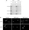
A, Immunoblot analysis with an antibody against human DBP in skin fibroblasts from one control subject, two DBP type I–deficient patients, and one patient with mild DBP type II deficiency that were cultured at 30°C and 40°C for 7 d. The arrowheads indicate the 79-kDa full-length protein, the 45-kDa enoyl-CoA hydratase plus sterol carrier protein 2–like units, and the 35-kDa 3-hydroxyacyl-CoA dehydrogenase unit. At 40°C, the 35-kDa band of DBP is absent in the fibroblasts of the patient with mild type II deficiency. B, DBP immunofluorescence microscopy in fibroblasts from a control subject, a DBP type I–deficient patient, and a patient with mild type II deficiency that were cultured at 30°C and 40°C for 7 d prior to immunofluorescence. At 30°C, the fibroblasts of the patient with mild type II deficiency show a normal punctate pattern of peroxisomes, like those of the control subject, whereas, at 40°C, the staining is negative.

References

Web Resources

    1. Online Mendelian Inheritance in Man (OMIM), http://www.ncbi.nlm.nih.gov/Omim/ (for DBP deficiency)
    1. Protein Data Bank (PDB), http://www.rcsb.org (for entries 1GZ6, 1S9C, 1IKT, and 1ZBQ)

References

    1. Adamski J, Carstensen J, Husen B, Kaufmann M, de Launoit Y, Leenders F, Markus M, Jungblut PW (1996) New 17 beta-hydroxysteroid dehydrogenases: molecular and cell biology of the type IV porcine and human enzymes. Ann N Y Acad Sci 784:124–136 - PubMed
    1. Adamski J, Normand T, Leenders F, Monte D, Begue A, Stehelin D, Jungblut PW, de Launoit Y (1995) Molecular cloning of a novel widely expressed human 80 kDa 17 beta-hydroxysteroid dehydrogenase IV. Biochem J 311:437–443 - PMC - PubMed
    1. Berman HM, Westbrook J, Feng Z, Gilliland G, Bhat TN, Weissig H, Shindyalov IN, Bourne PE (2000) The Protein Data Bank. Nucleic Acids Res 28:235–24210.1093/nar/28.1.235 - DOI - PMC - PubMed
    1. Bootsma AH, Overmars H, van Rooij A, van Lint AE, Wanders RJ, van Gennip AH, Vreken P (1999) Rapid analysis of conjugated bile acids in plasma using electrospray tandem mass spectrometry: application for selective screening of peroxisomal disorders. J Inherit Metab Dis 22:307–31010.1023/A:1005543802724 - DOI - PubMed
    1. Dieuaide-Noubhani M, Asselberghs S, Mannaerts GP, Van Veldhoven PP (1997) Evidence that multifunctional protein 2, and not multifunctional protein 1, is involved in the peroxisomal beta-oxidation of pristanic acid. Biochem J 325:367–373 - PMC - PubMed

Publication types

MeSH terms