Review
. 2005 Dec 31;46(6):737-49.
doi: 10.3349/ymj.2005.46.6.737.

Anatomic basis of sharp pelvic dissection for curative resection of rectal cancer

Affiliations
Review

Anatomic basis of sharp pelvic dissection for curative resection of rectal cancer

Nam Kyu Kim. Yonsei Med J. .

Abstract

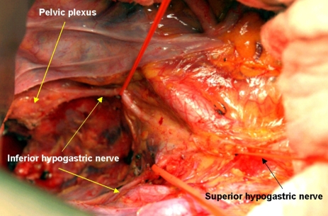
The optimal goals in the surgical treatment of rectal cancer are curative resection, anal sphincter preservation, and preservation of sexual and voiding functions. The quality of complete resection of rectal cancer and the surrounding mesorectum can determine the prognosis of patients and their quality of life. With the emergence of total mesorectal excision in the field of rectal cancer surgery, anatomical sharp pelvic dissection has been emphasized to achieve these therapeutic goals. In the past, the rates of local recurrence and sexual/voiding dysfunction have been high. However, with sharp pelvic dissection based on the pelvic anatomy, local recurrence has decreased to less than 10%, and the preservation rate of sexual and voiding function is high. Improved surgical techniques have created much interest in the surgical anatomy related to curative rectal cancer surgery, with particular focus on the fascial planes and nerve plexuses and their relationship to the surgical planes of dissection. A complete understanding of rectum anatomy and the adjacent pelvic organs are essential for colorectal surgeons who want optimal oncologic outcomes and safety in the surgical treatment of rectal cancer.


Figures

Fig. 1
Fig. 1
Cadaveric dissection of hemisectioned pelvis. (A) Presacral fascia covers the presacral vein over the sacrum. (B) The fascia picked up by the forceps is the rectal proper fascia enveloping the mesorectum and the rectum.
Fig. 2
Fig. 2
Cadaveric dissection of hemisectioned pelvis; the retrorectal space. The rectosacral fascia is noted in the retrorectal space at the level of 4th sacrum when dissection proceeds along the rectal proper fascial plane.
Fig. 3
Fig. 3
(A) The yellow line on the axial view of pelvic MRI is a imaginary line of sharp pelvic dissection along the rectal proper fascia (arrow). (B) A dotted line in schematic axial view is the loose areolar tissue plane between the rectal proper fascia and the parietal pelvic fascia. This line is the practical surgical dissection plane and can preserve the pelvic autonomic nerve without damaging to the mesorectum. The pelvic plexus is close contact with the fascia enveloping the mesorectum.
Fig. 4
Fig. 4
The mesorectum is well developed at the posterolateral side of the rectum. The mesorectum is tapered down and it ended 2-3 cm above the level of the levator ani muscle.
Fig. 5
Fig. 5
(A) The rectal proper fascia is adhesed to the mesh like pelvic plexus at the lateral pelvic wall. (B) The fine branches from pelvic plexus enter the rectal wall. The rectum was attached to the lateral pelvic wall by adhesed pelvic plexus.
Fig. 6
Fig. 6
(A) Midline of posterior side sacrum was divided and component of the levator ani muscle was shown. (B) U-shaped puborectalis muscle was shown around the rectum. These levator ani muscle must be cut off from its insertion site.
Fig. 7
Fig. 7
(A) Axial view of MR image shows a fine linear hypodense structure along the visceral pelvic fascia enveloping the mesorectum. (B) Coronal view of MR image shows a metastatic lymph node was located at close to the imaginary dissection line, especially the insertion site of the levator ani muscle.
Fig. 8
Fig. 8
Cadaveric dissection on hemisectioned pelvis show the inferior hypogastric nerve descend into the pelvic cavity and meet sacral parasympathetic nerve arising from S2th, 3th, 4th foramen nearby the piriformis muscle. The inferior hypogastric nerve form the pelvic plexus at the lateral pelvic wall after merging the sacral parasympathetic nerves. Nerve bundles from pelvic plexus go to the genitourinary organ along the seminal vesicle in male.
Fig. 9
Fig. 9
On operative field, bifurcation of the superior hypogastric nerve was noted at the aortic bifurcation. The inferior hypogastric nerve descends along the pelvic side wall. The pelvic plexus forms after merging with the sacral parasympathetic nerve.

Similar articles

Cited by

References

    1. NIH consensus conference. Adjuvant therapy for patients with colon and rectal cancer. JAMA. 1990;264:1444–1450. - PubMed
    1. Maurer CA, Z'Graggen K, Renzulli P, Schilling MK, Netzer P, Buchler MW. Total mesorectal excision preserves male genital function compared with conventional rectal cancer surgery. Br J Surg. 2001;88:1501–1505. - PubMed
    1. Heald RJ, Husband EM, Ryall RD. The mesorectum in rectal cancer surgery-the clue to pelvic recurrence? Br J Surg. 1982;69:613–616. - PubMed
    1. Heald RJ, Ryall RD. Recurrence and survival after total mesorectal excision for rectal cancer. Lancet. 1986;1:1479–1482. - PubMed
    1. MacFarlane JK, Ryall RD, Heald RJ. Mesorectal excision for rectal cancer. Lancet. 1993;341:457–460. - PubMed

Publication types