Review
. 2006 Apr;82(4):545-57.
doi: 10.1016/j.exer.2005.10.011. Epub 2006 Jan 4.

'What controls aqueous humour outflow resistance?'

Affiliations
Review

'What controls aqueous humour outflow resistance?'

Mark Johnson. Exp Eye Res. 2006 Apr.

Abstract

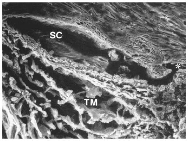
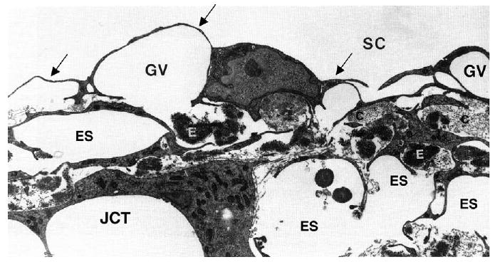
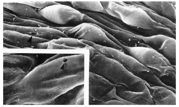
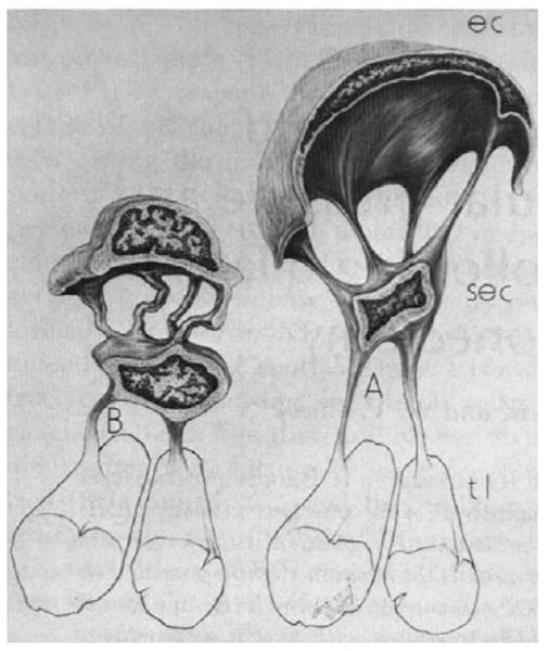
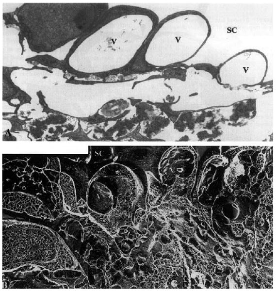
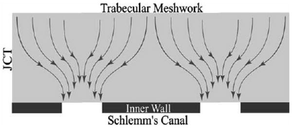
The bulk of aqueous humour outflow resistance is generated in or near the inner wall endothelium of Schlemm's canal in normal eyes, and probably also in glaucomatous eyes. Fluid flow through this region is controlled by the location of the giant vacuoles and pores found in cells of the endothelium of Schlemm's canal, but the flow resistance itself is more likely generated either in the extracellular matrix of the juxtacanalicular connective tissue or the basement membrane of Schlemm's canal. Future studies utilizing in vitro perfusion studies of inner wall endothelial cells may give insights into the process by which vacuoles and pores form in this unique endothelium and why inner wall pore density is greatly reduced in glaucoma.


Figures

Fig. 1
Fig. 1
Scanning electron micrograph of trabecular meshwork (TM) and Schlemm's canal (SC) from a human eye. Both regions are more expanded than they would be under physiological conditions. Asterisks shows a collecting channel (Freddo, 1993 (and revised 1999)). © 1993, Appleton and Lange.
Fig. 2
Fig. 2
Transmission electron micrograph of the inner wall of Schlemm's canal (SC) of a human eye, showing the endothelium (arrows) and giant vacuoles within this endothelial layer (GV). The region immediately below the inner wall and basement membrane is the juxtacanalicular connective tissue (JCT) and has many large, apparently empty spaces (ES). Collagen (C) and elastin (E) are apparent in the JCT along with fibroblastic-like cells. Magnification in source article ×89,100 (Gong et al., 1996). © 1996, Wiley–Liss, Inc.
Fig. 3
Fig. 3
Scanning electron micrograph of the inner wall of Schlemm's canal as viewed from within the canal. The bulging structures are giant vacuoles (and some nuclei). The insert shows an arrowhead pointing at a pore passing through one of the giant vacuoles (modified from (Allingham et al., 1992)). © 1992, Association for Research in Vision and Ophthalmology.
Fig. 4
Fig. 4
Schematic diagram showing change in configuration of cell (EC) of endothelial lining of Schlemm's canal going from low IOP (B) to high IOP (A). These cells attach to a second layer of cells (SEC) that in turn attach to the trabecular lamella (tl). (Johnstone, 1979). © 1979, Association for Research in Vision and Ophthalmology.
Fig. 5
Fig. 5
Enucleated human eye fixed by perfusion at 15 mmHg: (A) vacuoles (V) in the inner wall of Schlemm's canal (SC) in tissue prepared for TEM using conventional methods; notice the large open space in the region of the JCT immediately under these vacuoles; (B) the same region as seen in tissue prepared using QFDE; notice that while open spaces still exist under the vacuoles, a more complex and extensive extracellular matrix is seen (×4860) (Gong et al., 2002). © 2002, Elsevier.
Fig. 6
Fig. 6
(A) Schematic of use of micropipette to measure pressure in JCT as described by Maepea and Bill; (B) corrected schematic showing realistic size of micropipette in the JCT. Revised from (Maepea and Bill, 1992). © 1992, Elsevier.
Fig. 7
Fig. 7
Schematic of the ‘funnelling’ of aqueous humour through the JCT, toward a vacuole and pore that allows this fluid to pass through the inner wall endothelium (Overby et al., 2002). © 2002, Association for Research in Vision and Ophthalmology.
Fig. 8
Fig. 8
Pore density as a function of volume of fixative perfused through the outflow pathway of normal eyes (filled symbols) and eyes with POAG (open). Lines are best fit with outliers excluded. Details in Johnson et al. (2002). © 2002, Association for Research in Vision and Ophthalmology.

References

    1. Albelda SM, Sampson PM, et al. Permeability characteristics of cultured endothelial cell monolayers. J Appl Physiol. 1988;64:308–322. - PubMed
    1. Allingham RR, de Kater AW, et al. The relationship between pore density and outflow facility in human eyes. Invest Ophthalmol Vis Sci. 1992;33(5):1661–1669. - PubMed
    1. Alvarado JA, Yun AJ, et al. Juxtacanalicular tissue in primary open angle glaucoma and in nonglaucomatous normals. Arch Ophthalmol. 1986;104:1517–1528. - PubMed
    1. Alvarado JA, Murphy CG, et al. Effect of beta-adrenergic agonists on paracellular width and fluid flow across outflow pathway cells. Invest Ophthalmol Vis Sci. 1998;39:1813–1822. - PubMed
    1. Alvarado JA, Betanzos A, et al. Endothelia of Schlemm's canal and trabecular meshwork: distinct molecular, functional, and anatomic features. Am J Physiol Cell Physiol. 2004;286:C621–C634. - PubMed

Publication types