. 2006 Jan 5;49(1):11-6.
doi: 10.1016/j.neuron.2005.11.032.

A single vesicular glutamate transporter is sufficient to fill a synaptic vesicle

Affiliations

A single vesicular glutamate transporter is sufficient to fill a synaptic vesicle

Richard W Daniels et al. Neuron. .

Abstract

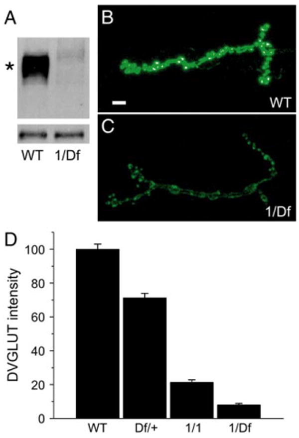
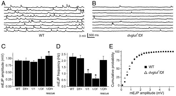
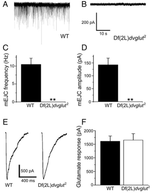
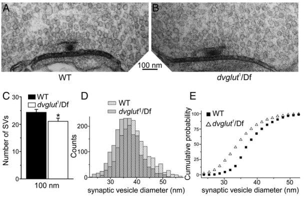
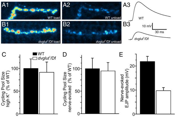
Quantal size is the postsynaptic response to the release of a single synaptic vesicle and is determined in part by the amount of transmitter within that vesicle. At glutamatergic synapses, the vesicular glutamate transporter (VGLUT) fills vesicles with glutamate. While elevated VGLUT expression increases quantal size, the minimum number of transporters required to fill a vesicle is unknown. In Drosophila DVGLUT mutants, reduced transporter levels lead to a dose-dependent reduction in the frequency of spontaneous quantal release with no change in quantal size. Quantal frequency is not limited by vesicle number or impaired exocytosis. This suggests that a single functional unit of transporter is both necessary and sufficient to fill a vesicle to completion and that vesicles without DVGLUT are empty. Consistent with the presence of empty vesicles, at dvglut mutant synapses synaptic vesicles are smaller, suggesting that vesicle filling and/or transporter level is an important determinant of vesicle size.


Figures

Figure 1
Figure 1. DVGLUT Protein Levels and Staining Are Reduced in dvglut Mutants
(A) Western blot of larval brain extracts from wild-type (WT) and dvglut1/Df(2L)dvglut2 (1/Df). Upper band (marked by an asterisk) is specific for DVGLUT, while lower band serves as a loading control. (B and C) Fluorescent staining for DVGLUT at muscle 4 of the third instar NMJ in control ([B], WT) and dvglut1/Df(2L)dvglut2 mutants ([C], 1/Df). Gain has been increased in both pictures to allow better visualization of the low levels of DVGLUT in the dvglut mutant. (D) α-DVGLUT staining intensity in wild-type controls (precise excision, labeled WT, n = 36), dvglut heterozygote (Df(2L)dvglut2/+, labeled Df/+, n = 13), homozygous hypomorph (dvglut1/dvglut1, labeled 1/1, n = 12), and the transheterozygote larvae (dvglut1/Df(2L)dvglut2, labeled 1/Df, n = 12). Data are shown as a percentage of wild-type staining intensity. All comparisons are significantly different with p < 0.001 (one-way ANOVA), error bars indicate SEM. Scale bar in panel (B) represents 5 μm.
Figure 2
Figure 2. mEJP Frequency but Not mEJP Amplitude Is Decreased in dvglut Mutants
Spontaneous activity was recorded intracellularly from muscle 6 in third instar larvae. Approximately 45 s of sample voltage recordings from control ([A], WT) and dvglut mutant ([B], dvglut1/Df(2L)dvglut2, labeled dvglut1/Df) larvae. (C) Histogram of average mEJP amplitudes for wild-type (WT, n = 30), dvglut heterozygotes (Df(2L)dvglut2/+, labeled Df/+, n = 10), homozygous hypomorphs (dvglut1/dvglut1, labeled 1/1, n = 10), and the transheterozygote (dvglut1/Df(2L)dvglut2, labeled 1/Df, n = 12) and rescue larvae (BG380 Gal4/+; dvglut1/Df(2L)dvglut2; UAS-DVGLUT/+, labeled 1/Df + rescue, n = 13). There is no significant difference in mEJP amplitude except in rescue larvae, which exhibit a slight increase in mini amplitude (p < 0.05, one-way ANOVA). (D) Histogram of average mEJP frequency for the same genotypes as in (B). A dose-dependent decrease in mEJP frequency is observed in dvglut1/dvglut1 (1/1) and dvglut1/Df(2L)dvglut2 (1/Df) larvae compared to wild-type. The difference between dvglut1/dvglut1 and dvglut1/Df(2L)dvglut2 is also significant, p < 0.05). (E) Cumulative probability distribution of minis from wild-type (WT, filled squares) and dvglut1/Df(2L)dvglut2 (1/Df, open triangles). The distributions are not significantly different by the K-S test (p > 0.2). *p < 0.05 versus wild-type. Error bars indicate SEM.
Figure 3
Figure 3. No Spontaneous Events Are Detected in dvglut Null Mutants Despite Normal Glutamate Responses
(A and B) Sample traces from wild-type (A) and dvglut null mutant embryos ([B], labeled Df(2L)dvglut2). (C and D) Average mEJC frequency (C) and amplitude (D) of spontaneous events in wild-type embryos. No events were detected in dvglut null mutants. (E and F) Glutamate receptors are present and respond equally to pressure ejection of glutamate. Sample individual current traces are shown in (E), and average response is shown in (F) (for wild-type [WT], n = 8, for Df(2L)dvglut2, n = 9, p > 0.8). Data are presented as mean ± SEM, **p < 0.01, Student's t test.
Figure 4
Figure 4. Synaptic Vesicles Are Abundant but Smaller on Average in dvglut Mutants
(A and B) Sample micrographs of active zones from control ([A], labeled WT) and mutant dvglut1/Df(2L)dvglut2 larvae ([B], labeled dvglut1/Df). (C) Synaptic vesicles within 300 nm of active zones were counted in 100 nm bins and the cumulative number of vesicles present within 100 nm of the active zone is shown for wild-type (filled bars, n = 43 active zones) and dvglut mutants (open bars, n = 39 active zones). Error bars indicate SEM. (D) Frequency histogram of synaptic vesicle outer diameters measured from EM sections of NMJs from wild-type (shaded bars, WT) and dvglut mutants (dvglut1/Df(2L)dvglut2, hatched bars). (E) Cumulative probability plot of the data in (D), with wild-type data (WT, black squares) and dvglut mutant data (dvglut1/Df, open triangles), n = 1840 vesicles for each genotype. The population of vesicle diameters is significantly shifted to smaller sizes in the dvglut mutant (K-S test, p < 0.05).
Figure 5
Figure 5. Cycling Vesicle Pool Is Unchanged in dvglut Hypomorphs, Despite Decreased EJP Amplitude
Pseudocolor image of wild-type (A1) and mutant (B1) synaptic boutons loaded with FM 1-43. (A2) and (B2) show the same region after unloading by nerve-evoked stimulation in wild-type and mutants, respectively. Intracellular recordings of nerve-evoked potentials are decreased in dvglut mutants. Sample traces from wild-type (A3) and mutant (B3) cells. (C) There is no difference in the optically determined cycling pool size after loading in 90 mM K+ saline and subsequently unloading in 90 mM K+ saline in wild-type (WT) and mutant (dvglut1/Df) larvae. (D) There is no difference in the optically determined cycling pool size after loading in 90 mM K+ saline and unloading by segmental nerve stimulation in normal saline in wild-type (WT) and mutant (dvglut1/Df) larvae. (E) Average nerve-evoked EJP amplitude is decreased in dvglut mutants (dvglut1/Df) compared to wild-type (WT) larvae. Error bars indicate SEM.

Comment in

  • Vesicular monogamy?
    Krantz DE. Krantz DE. Neuron. 2006 Jan 5;49(1):1-2. doi: 10.1016/j.neuron.2005.12.013. Neuron. 2006. PMID: 16387631

Similar articles

Cited by

References

    1. Boulland JL, Qureshi T, Seal RP, Rafiki A, Gunderson V, Bergersen LH, Fremeau RT, Jr, Edwards RH, Storm-Mathisen J, Chaudhry FA. Expression of the vesicular glutamate transporters during development indicates the widespread corelease of multiple neurotransmitters. J Comp Neurol. 2004;480:264–280. - PubMed
    1. Bruns D, Riedel D, Klingauf J, Jahn R. Quantal release of serotonin. Neuron. 2000;28:205–220. - PubMed
    1. Colliver TL, Pyott SJ, Achalabun M, Ewing AG. VMAT-mediated changes in quantal size and vesicular volume. J Neurosci. 2000;20:5276–5282. - PMC - PubMed
    1. Daniels RW, Collins CA, Gelfand MV, Dant J, Brooks ES, Krantz DE, DiAntonio A. Increased expression of the Drosophila vesicular glutamate transporter leads to excess glutamate release and a compensatory decrease in quantal content. J Neurosci. 2004;24:10466–10474. - PMC - PubMed
    1. Featherstone DE, Rushton EM, Hilderbrand-Chae M, Phillips AM, Jackson FR, Broadie K. Presynaptic glutamic acid decarboxylase is required for induction of the postsynaptic receptor field at a glutamatergic synapse. Neuron. 2000;27:71–84. - PubMed

Publication types

Substances