. 2006 Mar 15;90(6):1878-94.
doi: 10.1529/biophysj.105.071241. Epub 2005 Dec 30.

Dissecting the contribution of diffusion and interactions to the mobility of nuclear proteins

Affiliations

Dissecting the contribution of diffusion and interactions to the mobility of nuclear proteins

Joël Beaudouin et al. Biophys J. .

Abstract

Quantitative characterization of protein interactions under physiological conditions is vital for systems biology. Fluorescence photobleaching/activation experiments of GFP-tagged proteins are frequently used for this purpose, but robust analysis methods to extract physicochemical parameters from such data are lacking. Here, we implemented a reaction-diffusion model to determine the contributions of protein interaction and diffusion on fluorescence redistribution. The model was validated and applied to five chromatin-interacting proteins probed by photoactivation in living cells. We found that very transient interactions are common for chromatin proteins. Their observed mobility was limited by the amount of free protein available for diffusion but not by the short residence time of the bound proteins. Individual proteins thus locally scan chromatin for binding sites, rather than diffusing globally before rebinding at random nuclear positions. By taking the real cellular geometry and the inhomogeneous distribution of binding sites into account, our model provides a general framework to analyze the mobility of fluorescently tagged factors. Furthermore, it defines the experimental limitations of fluorescence perturbation experiments and highlights the need for complementary methods to measure transient biochemical interactions in living cells.


Figures

FIGURE 1
FIGURE 1
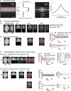
Test for diffusion-limited mobility NRK cells expressing PAGFP transiently (A), H2B-PAGFP stably (B), and PAGFP-RCC1 transiently (C). The first image of each dataset shows protein steady-state distribution in the nucleus imaged at 413 or 405 nm at low laser power before photoactivation. In all cases, half of the nucleus was photoactivated (open rectangle on second frame of each dataset). The last image represent 80% of fluorescence redistribution compared to steady state. To measure intensity profiles, each dataset was cropped using cropping regions like the one represented on the last frame of PAGFP. For each protein intensity, profiles were measured along the long axis of the nucleus, averaged along the short axis and normalized with the profile in steady state to generate fluorescence profiles for each time point. Plots display fluorescence intensity versus distance along the nucleus. The insets for PAGFP and H2B-PAGFP show the same profiles normalized between 0 and 1: note that whereas normalized profiles do not change for H2B-PAGFP, they become smoother for PAGFP and for PAGFP-RCC1. Scale bars: 5 μm.
FIGURE 2
FIGURE 2
Modeling. (A) Finite difference approach of reaction-diffusion model. The nucleus, in this case an NRK cell expressing transiently PAGFP-SUV39H1, is discretized in cuboids (images). The reaction occurs in each cuboid (arrows “binding”) and exchange of free proteins occurs between the nearest-neighbors by diffusion (solid arrows). (B) Simulation of a photoactivation experiment in three dimensions starting from a cell transiently expressing PAGFP-SUV39H1. The three-dimensional sequence shows the simulated nucleus before perturbation (first image) and during fluorescence redistribution, from the top and the side (total intensity projection).
FIGURE 3
FIGURE 3
Two-dimensional simplification. (A) PAGFP-RCC1 fixed in a 30% acrylamide gel was photoactivated in the red region using a 40× iris objective with a numerical aperture fixed to 1.0, seen from the top and the side. Scale bar: 5 μm. The longitudinal profile shows the average intensity of the profile generated by photoactivation in the confocal section where photoactivation was focused. The red curves correspond to a fit of half of this profile with the error function. The axial intensity profile corresponds to the profile of illumination in depth along the arrow of the image. (B) Simulation of a photoactivation experiment in three dimensions, using the depth profile from panel A and a Gaussian radial PSF for the photoactivation profile (first row, total intensity top and side projection), and two-dimensional observation of the simulation, using the same depth profile and radial PSF as for the photoactivation profile (second row). The first images of each row represent the steady-state distribution of fluorescence and the following represent the fluorescence redistribution. The first plot shows the average fluorescence intensity over time of the six regions depicted on the last images of the two-dimensional sequence (circles) and the fit using the simplified two-dimensional model (solid curves). The second plot represents the residuals, <1% for the six regions, between the three-dimensional simulations and the two-dimensional fit. (C) Three-dimensional simulation with a higher percentage of free proteins, starting from cell stably expressing PAGFP-SUV39H1 (first row). The second plot in the first row is a zoom of the early phase of the first plot and shows the diffusion of the initial free pool of fluorescent proteins. The amplitude of this early phase is related to the amount of free proteins. The late phase visible on the first plot corresponds mostly to the kinetics of the interaction. In this case, the same regions as in B cannot be well fitted with the two-dimensional simplified model (second row) with residuals reaching 25%, but it improves drastically (see solid curves and residuals of the third row) and parameters are close to the three-dimensional situation when one uses only the two regions depicted on the image of the third row. Scale bars: 5 μm.
FIGURE 4
FIGURE 4
Parameter identifiability. (A) Fit and residuals for Fig. 3 B starting from two different fixed diffusion coefficients. It should be noted that the fits are almost similar, showing that in such a case the diffusion constant has to be determined separately to be able to estimate the other parameters. (B) PAGFP photoactivation. The nucleus was photoactivated (open region, first image) and imaged over time (first row). The intensities of the six regions depicted on the last image were plotted over time (upper plot, circles) and fitted (solid curves). Residuals are below 6% (lower plot). The sequence on the second row is the simulation using the parameter from the fit. Scale bar: 5 μm.
FIGURE 5
FIGURE 5
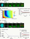
PAGFP-RCC1. The percentage of free molecules and only the lower limit of dissociation rate can be estimated. (A) Nucleus of NRK cell transiently expressing PAGFP-RCC1, acquired at 405 nm, low power, and 488 nm before photoactivation (first two images) and at 488 nm after activation (second row). The plots represent the average intensity over time of the regions depicted on the last image (circles) and the fit (solid curves, first plot), and the residuals (second plot). The simulation using the parameters from the fit is shown on the last image row. Scale bar: 5 μm. (B) Color-coded sum of the square of the residuals for different values of dissociation rates koff and percentage of free proteins. The black-cross fit on the parameter space corresponds to the fit in A. The regions with white boundaries correspond to the values of sum of residual squares that are less than the double of the one corresponding to the fit. (C) Same plots as in panel A, but using a instantaneous reaction model. Note that it is almost completely similar to panel A.
FIGURE 6
FIGURE 6
PAGFP-SUV39H1 and H1.1PAGFP. Different timescales but same conclusions as for PAGFP-RCC1; only the lower limit of dissociation rate can be estimated. (A) Nucleus of an NRK cell stably expressing PAGFP-SUV39H1, acquired at 405 nm, low power, before activation (first image) and at 488 nm before and after activation of half of the nucleus (open region, second image). (B) Parameter space as in Fig. 5 B. (C) Instantaneous reaction model for PAGFP-SUV39H1, almost similar to a reaction-diffusion model (not shown). (D) NRK cell stably expressing H1.1-PAGFP. Images as in A. (E) The parameter space represents the sum of the squares of the residuals for different values of dissociation rates koff, the percentage of free proteins being fixed to the value given by the fit. The horizontal dashed line corresponds to the double of the minimum of this sum, giving the lower limit of dissociation rate 0.017 s−1 depicted on the plot. (F) Instantaneous reaction model for H1.1-PAGFP, similar to a reaction-diffusion model (not shown). Scale bars: 5 μm.
FIGURE 7
FIGURE 7
PAGFP-SUV39H1-H320R case. Free pool of 35% and residence time of 210 s. Nucleus of NRK cell stably expressing the hyperactive PAGFP-SUV39H1-H320R, acquired at 488 nm. Contrary to the other cases, the steady-state distribution was not measured at 405 nm as the signal was too low. The intensities of the two regions depicted on the last image are plotted (circles) and fitted (solid curves) as in Fig. 3 C. The second plot correspond to a zoom of the early part of the first plot.
FIGURE 8
FIGURE 8
Estimation of dissociation rates. Experimental and theoretical limits. Positions of the different constructs on the diagram of fraction of free proteins versus dissociation rates. The curve between the shaded and the unshaded regions corresponds to the limit of dissociation rates that can be estimated, with a tolerance of 5%, determined from the comparison between reaction-diffusion and instantaneous reaction models. The shaded region corresponds to the space where the dissociation rate cannot be estimated. It should be noted that although the dissociation rates of H1.1-PAGFP, PAGFP-RCC1, and PAGFP-SUV39H1 cannot be determined, their limit is outside the shaded region, likely because this limit also takes into account data noise and systematic errors.

References

    1. Houtsmuller, A. B., S. Rademakers, A. L. Nigg, D. Hoogstraten, J. H. Hoeijmakers, and W. Vermeulen. 1999. Action of DNA repair endonuclease ERCC1/XPF in living cells. Science. 284:958–961. - PubMed
    1. Phair, R. D., and T. Misteli. 2000. High mobility of proteins in the mammalian cell nucleus. Nature. 404:604–609. - PubMed
    1. Misteli, T., A. Gunjan, R. Hock, M. Bustin, and D. T. Brown. 2000. Dynamic binding of histone H1 to chromatin in living cells. Nature. 408:877–881. - PubMed
    1. McNally, J. G., W. G. Muller, D. Walker, R. Wolford, and G. L. Hager. 2000. The glucocorticoid receptor: rapid exchange with regulatory sites in living cells. Science. 287:1262–1265. - PubMed
    1. Bubulya, P. A., and D. L. Spector. 2004. On the movements of nuclear components in living cells. Exp. Cell Res. 296:4–11. - PubMed

Substances

LinkOut - more resources